Protein by Candlelight, Grapes beneath the Scalpel
Urine and pus. Two small pools of fluid, two very different rooms. One belonged to a dim ward in 1827 London, where Richard Bright warmed a…
Protein by Candlelight, Grapes beneath the Scalpel
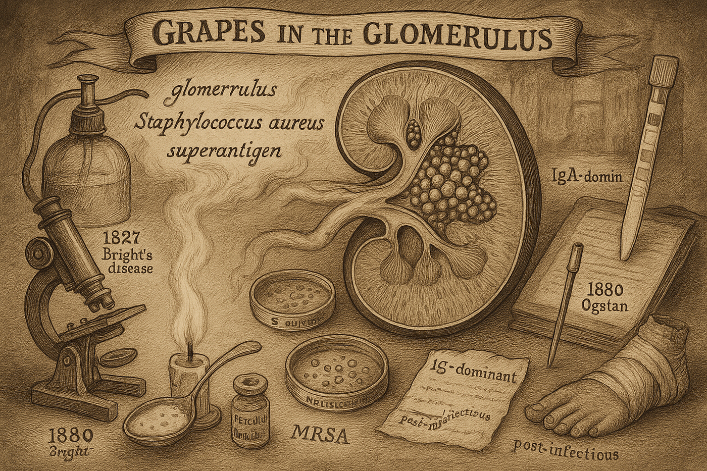
Urine and pus. Two small pools of fluid, two very different rooms. One belonged to a dim ward in 1827 London, where Richard Bright warmed a spoonful of cloudy urine over a candle. The other lay in an Aberdeen operating theater in 1880, where Alexander Ogston focused his microscope on a drop of pus. Neither man could have guessed that their observations would form the opening moves in a medical pursuit that would take more than a century to resolve: how an infection on the skin could ignite a storm in the kidney.
What Bright saw was ordinary to the eye yet remarkable by heat: the urine turned milky and clotted like egg drop soup. Protein was present, and it precipitated when warmed. What Ogston saw was elegant and lethal: clusters of tiny spheres gathered in bunches, like translucent grapes. He called them Staphylococcus, from the Greek for grape and berry. Two clues, two cities, two lines that would not cross for nearly a hundred years.
Bright’s candle
To understand where the chase began, we need to meet Richard Bright. He was not only a physician but a restless observer, a man who sketched landscapes, studied rocks, and volunteered among the wreckage of war. The gift that made him dangerous to disease was not a new machine but a habit of mind. He touched, listened, percussed, and peered. He compared what bodies said in life with what organs confessed after death.
At the time, dropsy was a baggy diagnosis for swelling. Hearts, livers, kidneys, and bad luck could all be blamed; the distinctions were muddy. Bright changed that by building a clinical bridge: correlate the patient’s symptoms with postmortem anatomy. He spent hours shuttling between wards and autopsy tables, assembling a pattern. Swollen patients tended to have heat-precipitable protein in their urine and granular, damaged kidneys at autopsy. In 1827, he published a set of cases that tied the triad together and gave the world a sharper concept: Bright’s disease. The insight was larger than a name. He took a systemic symptom and traced it to a single failing organ, sketching the early borders of what would later be called nephrology.
Years before the blood pressure cuff, Bright’s fingertips detected a hard, unyielding pulse and enlarged hearts in those same patients. He sensed an alliance between hypertension and kidney injury without instruments to prove it. His candle did not simply reveal a biochemical trick. It lit a philosophy: simple tools in prepared hands can answer deep questions.
Grapes under the knife
While Bright refined diagnosis in London, surgery elsewhere was still a brutal gamble. Infection was the real killer, and the stink of carbolic acid became the perfume of progress when Joseph Lister brought antisepsis into the theater. Ogston, a surgeon in Aberdeen, admired Lister. Yet he wanted more than safer technique. He wanted to see the enemy.
In 1880 he drew a bead of pus from an abscess and set it on a slide. Through his lens, two microcosms emerged: chains of spheres already known as streptococci, and grape-like clusters that had no name. Ogston baptized them Staphylococcus and then did the hard work of proof. He isolated the clusters and injected them into animals, reproducing abscesses. Causality, not coincidence.
A few years later, Friedrich Julius Rosenbach cultivated these organisms and noticed their colors: some colonies gleamed gold, others white. The golden strain took the name Staphylococcus aureus and eventually took center stage as one of medicine’s most resourceful villains. The scientific drama, though, had its irony. Lister, the father of antisepsis, initially doubted Ogston’s precise microbe-centric view, preferring a more general theory of putrefaction. The field was pivoting from vague miasmas to specific culprits. Modern microbiology was learning to write the disease story with proper nouns.
The plot thickened in the antibiotic era. Penicillin felled S. aureus, then resistance rose. Methicillin arrived in 1959, and methicillin-resistant S. aureus announced itself by 1961. The duel has not ended. The grapes under Ogston’s scalpel were not just pretty; they were adaptive.
A century of silence between clues
For decades, Bright’s candle and Ogston’s grapes lived in separate constellations. Kidney doctors mapped the shapes of diseased glomeruli. Microbiologists cataloged infections and invented new drugs. A clinician treating nephritis rarely imagined a connection to a healing wound or a crusted boil. How did the lines finally cross?
Technology brought the threads together. Two inventions mattered. First, the democratization of urinalysis: dipsticks turned the old spoon-and-candle trick into a pocket-sized, seconds-long assay. Protein and blood could be flagged at the bedside as routinely as checking a pulse. Second, the renal biopsy: a needle to the living kidney, a path to histology without waiting for death. With light microscopy, immunofluorescence, and eventually electron microscopy, pathologists could watch the battle inside the glomerulus in real time.
What they saw surprised them. The kidney was not a battlefield littered with bacteria. It was a battlefield littered with our own collateral damage.
Friendly fire
The immune system is a disciplined army when it recognizes the right foe. Antibodies bind to antigens, immune complexes form, cleanup crews arrive, and the blood runs clean again. When the fight is furious, though, debris accumulates. Excess immune complexes circulate and lodge where the blood is filtered most finely: the glomerular capillary bed. There, the complexes spark complement activation and summon inflammatory cells. The filter swells and leaks. Protein spills. Red cells escape. The urine tells the story long before the patient learns the words.
But Staphylococcus aureus does more than provoke the usual skirmish. It carries a cheat code known as a superantigen. Instead of politely presenting itself to a small fraction of T cells that fit its shape, it forces a mass rally. It bypasses the checks and floods the field, activating an enormous slice of the T cell population at once. Cytokines surge. B cells fire indiscriminately. Antibodies bloom in messy profusion. Immune complexes rise like silt in a storm, and the kidney’s sieve clogs with the fallout.
In this light, staphylococcal glomerulonephritis is not a direct invasion of the kidney by bacteria. It is arson by proxy. The bug lights the match, but our own overzealous firefighters drench the house until the roof caves in.
The modern face of a golden foe
If you trained on the classic story of post-streptococcal glomerulonephritis, you learned to expect a child who had a sore throat or impetigo a couple of weeks ago, now swollen and producing cola-colored urine. The infection is over, the immune response lingers, and the kidney suffers late.
Staphylococcus-associated glomerulonephritis is different. It tends to find older adults, often men, carrying the burdens of diabetes, liver disease, alcoholism, or devices that breach the skin and enter the bloodstream. The infection is not past; it is ongoing. The kidney injury blooms while the battle with S. aureus rages somewhere in the body: a foot ulcer, osteomyelitis, pneumonia, or an infected line.
In a Taiwanese ward, the story might look like this. Mr. Chen is 65, with long-standing diabetes. He is admitted for a stubborn foot infection. A week into treatment, his urine darkens and his legs swell. His creatinine leaps. The dipstick lights up for protein and blood. Biopsy shows glomeruli thick with inflammation and immune deposits that glow for IgA on immunofluorescence. That immunofluorescence pattern can masquerade as primary IgA nephropathy, but the surrounding context gives it away: the infection is active and the organism is often staphylococcus. This is SAGN.
Treatment starts by hunting and eradicating the source. Antibiotics tailored to the organism, surgical drainage if needed, and removal of infected hardware when present. Whether to add steroids is a careful calculus. In some cases, tamping down the immune fire helps preserve function; in others, it risks pouring gasoline on the infection. Even with the best choices, the outlook is guarded. Resistant strains like MRSA complicate the path, and a notable fraction of patients do not return to their prior kidney function. Some require dialysis. Mortality is higher than in the post-streptococcal disease of childhood memory.
There is a quiet paradox at work. Modern medicine keeps people with complex illnesses alive longer and more comfortably. We insert catheters, place grafts, and administer life-extending therapies. We also build a larger, more vulnerable population in whom S. aureus finds new doors. Progress can carry its own shadows.
From history back to the clinic
Return to the two rooms. The candle that turned urine cloudy is, in a way, the ancestor of every dipstick on every nurse’s cart. The grape-like clusters under Ogston’s lens live on in every culture plate and every antibiogram. The biopsy needle and the fluorescent microscope finally brought their clues to the same table, and immunology told us why the story hurt so much: a wily bacterium weaponizing our own responses.
For working clinicians, this is not a museum piece. When a biopsy reads IgA-dominant immune complex deposition, the right next step is not reflexive steroid dosing but detective work. Where might a staphylococcal infection be hiding. A healing wound that never quite healed. A line that has lingered too long. A spine that aches with an unhelpful fever. Our tools grow more powerful each year, but they still require Bright’s curiosity and Ogston’s insistence on seeing the culprit. The deeper lesson is durable: the organ, the organism, and the host response are parts of one narrative. We honor patients when we read all three.
Some stories begin with a single flame. This one still burns in our wards and labs. Every time protein fogs a dipstick and a pathologist’s slide glows with IgA, the old clues whisper. Be thorough. Look for the grapes. Treat the infection. Respect the kidney’s quiet testimony.
Keywords: staphylococcus kidney; immune complex storm; Bright’s candle test; MRSA and SAGN; clinical detective work
References
- Bright R. Reports of medical cases, selected with a view of illustrating the symptoms and cure of diseases by a reference to morbid anatomy. Volume 1. London: Longman, Rees, Orme, Brown, and Green; 1827.
- Ogston A. On Abscesses. Rev Infect Dis. 1984;6(1):122–8.
- Rosenbach FJ. Microorganismen bei den Wund-Infections-Krankheiten des Menschen. Wiesbaden: J.F. Bergmann; 1884.
- Cameron JS. Richard Bright, MD (1789–1858): the man, his life and his legacy. Kidney Int. 2002;61(6):2278–84.
- Orenstein A. The Discovery and Naming of Staphylococcus aureus. Antimicrobe.org. [Internet]. [cited 2024 May 20]. Available from: http://www.antimicrobe.org/h04c.files/history/S-aureus.pdf
- Chambers HF, Deleo FR. Waves of resistance: Staphylococcus aureus in the antibiotic era. Nat Rev Microbiol. 2009;7(9):629–41.
- Iversen P, Brun C. Aspiration biopsy of the kidney. Am J Med. 1951;11(3):324–30.
- Cameron JS. The introduction of renal biopsy into nephrology from 1901 to 1961: a paradigm of the forming of nephrology by technology. Am J Nephrol. 1997;17(3–4):347–58.
- Berger J, Hinglais N. Les dépôts intercapillaires d’IgA-IgG [Intercapillary deposits of IgA-IgG]. J Urol Nephrol (Paris). 1968;74(9):694–5. French.
- Lerner RA, Glassock RJ, Dixon FJ. The role of anti-glomerular basement membrane antibody in the pathogenesis of human glomerulonephritis. J Exp Med. 1967;126(6):989–1004.
- Schlievert PM, Shands KN, Dan BB, Schmid GP, Nishimura RD. Identification and characterization of an exotoxin from Staphylococcus aureus associated with toxic-shock syndrome. J Infect Dis. 1981;143(4):509–16.
- Marrack P, Kappler J. The staphylococcal enterotoxins and their relatives. Science. 1990;248(4956):705–11.
- Koyama A, Kobayashi M, Yamaguchi N, Yamagata K, Takano K, Nakajima M, et al. Glomerulonephritis associated with methicillin-resistant Staphylococcus aureus infection: a possible role of bacterial superantigen. Clin Nephrol. 1995;43(4):205–10.
- Satoskar AA, Nadasdy G, Plaza JA, Sedmak D, Shidham G, Hebert L, et al. Staphylococcus infection-associated glomerulonephritis mimicking IgA nephropathy. Clin J Am Soc Nephrol. 2006;1(6):1179–86.
- Nasr SH, Markowitz GS, Whelan JD, D’Agati VD. IgA-dominant acute poststaphylococcal glomerulonephritis. Hum Pathol. 2003;34(12):1235–41.
- Haas M. IgA-dominant infection-associated glomerulonephritis: a distinct clinicopathologic entity. Kidney Int. 2017;92(4):784–787.
- Nadasdy T, Hebert LA. Infection-related glomerulonephritis: understanding mechanisms. Semin Nephrol. 2011;31(4):369–75.
- Wang S, Bu R, Zhang Q, Wu J, Wang L, Liu X, et al. Clinical, Pathological, and Prognostic Characteristics of Glomerulonephritis Related to Staphylococcal Infection. Medicine (Baltimore). 2016;95(19):e3618.
메타데이터
- post_id
- 5301b84feb60
- slug
- protein-by-candlelight-grapes-beneath-the-scalpel-5301b84feb60
- url
- https://medium.com/medical-stories/protein-by-candlelight-grapes-beneath-the-scalpel-5301b84feb60
- canonical_url
- https://medium.com/medical-stories/protein-by-candlelight-grapes-beneath-the-scalpel-5301b84feb60
- author_url
- https://medium.com/@critnephro
- status
- ok
- fetched_at
- 2026-06-13 00:08:42