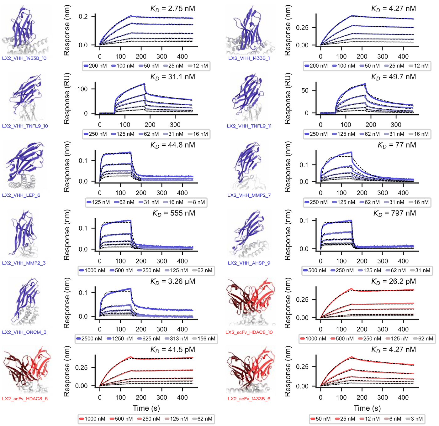
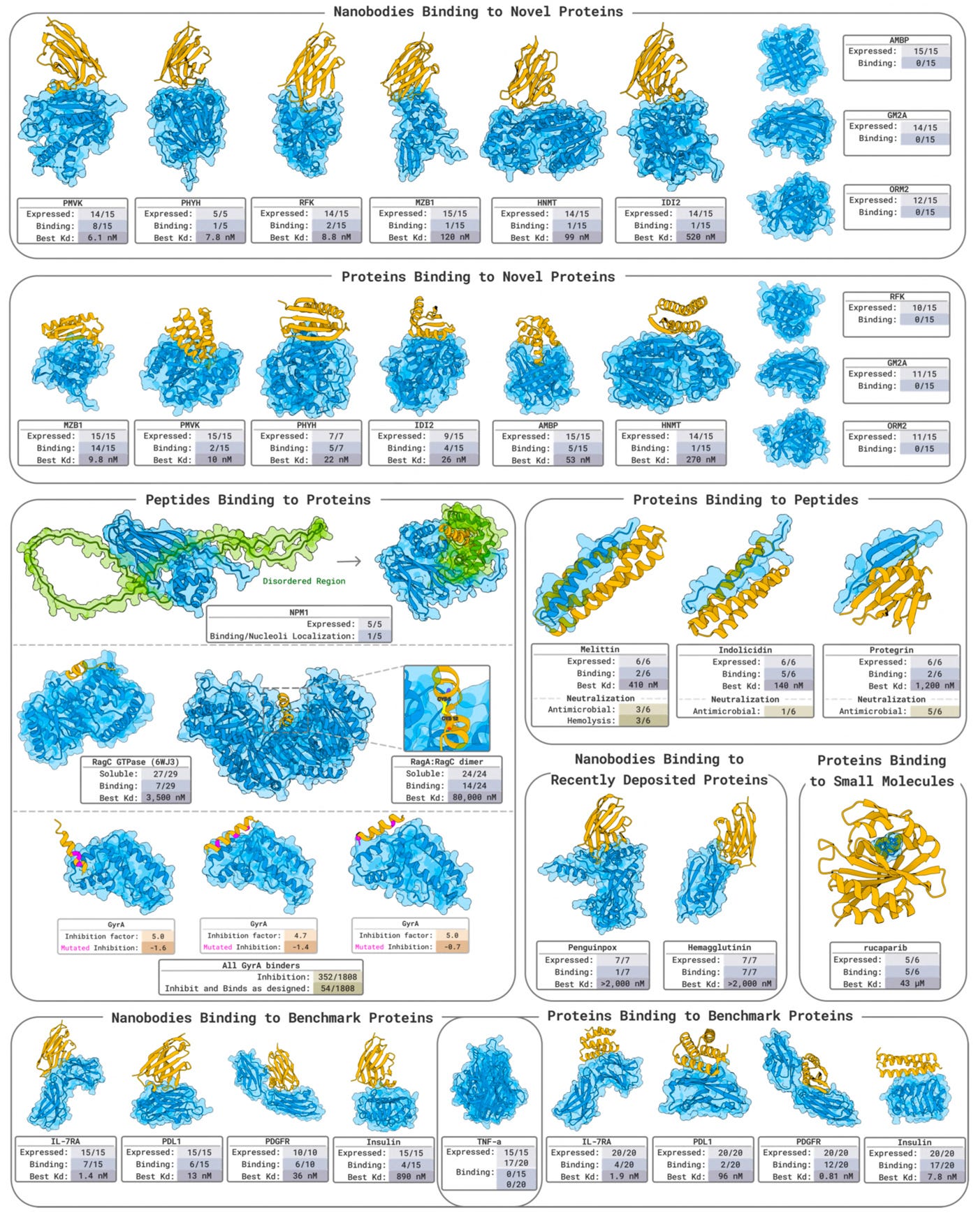
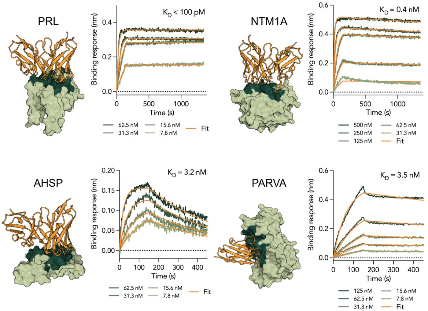
Origin-1: Absci’s De Novo Antibody Design Platform
Micromolar hits with structural validation but therapeutic gaps.
Origin-1: Absci’s De Novo Antibody Design Platform
Disclosure: I previously worked at Absci.
Absci published Origin-1, a computational platform for de novo antibody design against epitopes lacking prior structural data. In this post, I summarize what the paper reports and identify gaps in the experimental validation.
Platform Overview
Origin-1 comprises three components:
- AbsciDiff: Diffusion-based structure generation, fine-tuned from Boltz-1. The model generates antibody-antigen complexes conditioned on epitope specification. Key modifications include epitope conditioning through feature masking, a sequence hypothesis module, and template support. Training used 10,045 antibody-antigen structures from PDB with temporal and sequence homology filters.
- IgDesign2: CDR sequence design model with a GNN encoder, causal transformer decoder, and IgBert³ refinement. Takes structural predictions from AbsciDiff and generates CDR sequences compatible with the predicted binding geometry.
- AbsciBind: Scoring protocol combining ipTM and a custom Antibody-Aligned ipTM metric. Both metrics were developed by Absci for this paper. The score is derived from AF_Unmasked¹ with modifications: amino acid tokens retained, side chains masked except Cβ, and inter-chain template distances enabled. Designs with AbsciBind Score ≥0.5 were considered high-confidence.
Three-stage filtering pipeline:
Wide Structure Search: 3,360 design specifications sampled (combinations of frameworks, CDR lengths, epitope residues). AbsciDiff generated 80,640 structures (24 diffusion samples per specification), selected 10,080 for scoring. IgDesign2 generated one sequence per structure. Top 10% by AbsciBind Antibody-Aligned ipTM Score advanced (1,008 design specifications, oversampled 10× to 10,080 specifications for Deep Search).
Deep Structure Search: 10,080 design specifications from Wide Search. AbsciDiff generated 241,920 structures, selected 30,240 for scoring. Pooled with Wide Search structures (40,320 total). Filtered by L-RMSD <5Å between designed and predicted structures. Top 500 structures by Antibody-Aligned ipTM Score and Intersection Score advanced to Sequence Search. Structural diversity enforced through agglomerative clustering (maximum 30% from single cluster).
Sequence Search: 320 sequences generated per structure (500 structures = 160,000 sequences). Filtered for sequence liabilities and deduplicated. Top 20 sequences per structure scored with AbsciBind. Final selection: 95 designs per target, balancing AbsciBind Score with diversity constraints (maximum 3 replicates per HCDR3 sequence, maximum 30% from single structural cluster).
Per target totals: ~322,000 structures generated, ~40,000 structures scored, ~200,000 sequences generated, ~50,000 sequences scored with AbsciBind.
Targets, Hit Rates, and Hit Affinities
Ten human protein targets were selected based on: high structural resolution (<3.5 Å), few missing residues, commercial antigen availability, no existing antibody-antigen complex structures in PDB, and ≤60% sequence identity to any protein with a known complex.

* Paper text reports 9 total SPR hits (COL6A3: 3, AZGP1: 4, CHI3L2: 1, IL36RA: 1), but GitHub repository contains binding data for only 5 designs.
Hit affinities:
The paper states they got “9 total SPR hits” but only reports affinities for 5 binders. The GitHub repository confirms only 5 designs as binders (marked “True” in ‘de_novo_spr_data.csv’ and have SPR KD values reported in ‘lead_optimization_spr_data.csv’):
COL6A3: 1 hit, 1.94 µM.
AZGP1: 2 hits, 1.42 and 4.50 µM. No BLI confirmation data for the second hit.
CHI3L2: 1 hit, 3.46 µM. Failed BLI confirmation.
IL36RA: 1 hit, 6.07 µM. Failed BLI confirmation.
Structural Validation
Cryo-EM structures were solved for COL6A3 and AZGP1 complexes:
COL6A3 (3.0 Å resolution):
- All-atom global RMSD: 2.56 Å
- Interface RMSD: 0.96 Å
- DockQ²: 0.83
- CDR RMSDs: 0.661–1.486 Å
AZGP1 (3.1 Å resolution):
- All-atom global RMSD: 1.79 Å
- Interface RMSD: 1.35 Å
- DockQ: 0.73
- CDR RMSDs: 0.751–2.056 Å

COL6A3 binding validation and structural confirmation for the strongest de novo hit (1.9 µM parent affinity). (A) SPR sensorgrams show concentration-dependent binding to COL6A3 in mAb format with no cross-reactivity to off-targets. Note the biphasic kinetics. (B) BLI confirms binding in Fab format when antibody is immobilized; binding failed when antigen was immobilized (data not shown). (C) SEC shows complex formation between COL6A3 and Fab, though complex formation required Nanodaptor and Kappabulk stabilizers. (D) Cryo-EM structure at 3.0 Å resolution shows 0.96 Å interface RMSD between computational model (red/black/gray) and experimental structure (salmon), validating the predicted binding mode. (E) Individual CDR overlays demonstrate sub-2Å structural accuracy across all CDRs.

AZGP1 binding validation and structural confirmation for the strongest de novo hit (1.4 µM parent affinity). (A) SPR sensorgrams show concentration-dependent binding to AZGP1 in mAb format with no cross-reactivity to off-targets. (B) BLI confirms binding in Fab format when antibody is immobilized. Unlike COL6A3, AZGP1 designs also bound in the flipped orientation with antigen immobilized (data not shown in figure). (C) SEC shows complex formation between AZGP1 and Fab without requiring additional stabilizers. (D) Cryo-EM structure at 3.1 Å resolution shows 1.35 Å interface RMSD between computational model (red/black/gray) and experimental structure (salmon), validating the predicted binding mode. DockQ score of 0.73 indicates good model quality. (E) Individual CDR overlays demonstrate structural prediction accuracy across all six CDRs. Despite the structural validation, this design was flagged for multiple developability concerns: self-association (25.7 nM AC-SINS), polyreactivity against both insulin (15.8 RNS) and DNA (8.5 RNS), and hydrophobicity. The 1.4 µM affinity is the strongest in the paper but remains well below what would typically justify downstream therapeutic development.
The COL6A3 complex required Nanodaptor and Kappabulk stabilization for cryo-EM grid preparation. In BLI, COL6A3 showed binding only in the antibody-immobilized orientation; it failed to bind when antigen was immobilized.
CHI3L2 and IL36RA binders (including optimized variants with improved affinity and functional activity) do not have experimental structures.
Lead Optimization
Single-mutant libraries (94 variants + 1 parent) were designed using AbsciBind scores combined with ESM ensemble and AbLang2. The GitHub repository contains KD values for 354 optimization variants.
Optimization hit rates (variants showing actual improvement over parental molecules):
- AZGP1-A: 21/94 (22%)
- AZGP1-B: 28/94 (30%)
- IL36RA: 31/94 (33%)
- CHI3L2: 28/94 (30%)
- COL6A3: 24/94 (26%)
Of the 94 variants tested per parent, 22–33% showed improved affinity over the parental molecule. But of the 354 optimization variants with KD values, 324 remained above 1 µM. Nine variants reached sub-500 nM affinity, and just three broke 200 nM.
Computational Benchmark
AbsciGen was compared to a modified version of RFantibody. Modifications were required because RFantibody’s default coordinate initialization creates chain breaks for full-length antibodies, and public examples contained actual PDB loop coordinates.
Results using AbsciBind Score ≥0.5 threshold:
- AbsciGen: 28.37% of designs passed
- RFantibody: 1.49% of designs passed
AbsciBind Score is the average of ipTM and Antibody-Aligned ipTM, both metrics developed by Absci for this paper.
Methodological Transparency
The level of detail in this paper exceeds typical industry publications. The authors describe:
Model architecture and training:
- AbsciDiff’s modifications to Boltz-1 (feature masking, epitope conditioning, sequence hypothesis module, template support)
- IgDesign2’s architecture (GNN encoder, causal transformer decoder, IgBert refinement)
- Training data curation (AbData pipeline, 10,045 PDB structures, inclusion/exclusion criteria, temporal and sequence homology filters)
- Cropping strategy (512 residue maximum, Fv always included, constant domains excluded)
- Fine-tuning parameters (20 learning rate steps, batch size 64, 10 epochs)
Design generation pipeline:
- Three-stage filtering (Wide Structure Search => Deep Structure Search => Sequence Search)
- Design specification parameters (HCDR3 length 8–26, LCDR3 length 8–10, epitope subsampling 45–90%)
- 3360 design specifications sampled per target, yielding ~40,000 structures and ~170,000 sequences
- Framework selection rationale (trastuzumab, relatlimab, sotrovimab, and dupilumab to cover diverse germlines)
Epitope selection:
- Criteria for each target (surface geometry, curvature, hydrophobic patches, structured regions)
- Specific residues selected per epitope (Figure 6 in the paper)
- IL36RA epitope derived from structural homology to IL36γ small molecule binding site (PDB: 6P9E)
- Acknowledgment of AZGP1 PDB entry missed during selection
Scoring protocol:
- AbsciBind derivation from AF_Unmasked
- Modifications to AFM protocol (amino acid tokens retained, side chains masked except Cβ, inter-chain template distances enabled)
- Benchmark against six reference approaches with performance comparison
Experimental methods:
- SPR parameters (chip type, buffer composition, regeneration conditions, analysis software, exclusion criteria)
- BLI protocol (sensor types, baseline, association/dissociation times)
- Developability assays (methods for each assay, system suitability controls)
- Cryo-EM data collection and processing (microscope settings, software pipeline)
Fair Benchmark Against RFantibody
The authors identified problems with RFantibody that would have artificially inflated their comparative results:
- Chain breaks: Running RFantibody without modification produced chain breaks in predicted loop structures and glycine-dominated sequences from ProteinMPNN.
- Information leakage: The public RFantibody release includes actual loop coordinates from PDB entries in the provided framework files. Using these coordinates in a “de novo” benchmark would leak structural information.
- Initialization bias: RFantibody’s default coordinate initialization causes spatial biases to persist through the diffusion process.
Rather than using RFantibody out of the box (which would have made Origin-1 look better by comparison), the authors:
- Preprocessed structures to place CDR loop coordinates at the origin
- Applied idealized initialization based on loop dilation logic
- Documented the modifications in Supplement §7.3
- Showed results comparing original vs. modified RFantibody (Supplementary Figure 6)
The paper explicitly states this was done “to ensure a fair benchmark.” They could have published a comparison against the unmodified RFantibody showing larger performance gaps. Choosing to fix the competitor’s implementation before comparison is commendable.
The methods sections are detailed and the RFantibody benchmark was handled fairly: they fixed the coordinate initialization problem before comparison rather than capitalizing on a broken baseline. But detailed protocols for generating micromolar binders don’t make those binders therapeutically useful. Several gaps and inconsistencies make the interpretation of the results difficult.
Gap #1: Micromolar Affinities Have Limited Therapeutic Relevance
Parent design affinities from SPR:
- COL6A3: 1.94 µM
- AZGP1: 1.42 and 4.50 µM
- CHI3L2: 3.46 µM
- IL36RA: 6.07 µM
Best affinities after optimization:
- IL36RA: 89.4 nM
- COL6A3: 174 nM
- AZGP1: 193 nM
CHI3L2 and IL36RA hits showed SPR signals but could not be confirmed by BLI prior to optimization. The authors attributed this to low affinity (KD > 2 µM). However, the second AZGP1 hit (4.50 µM) is weaker than CHI3L2 (3.46 µM) yet was reportedly confirmed by BLI (though no data for this second hit appears in Figure 10).
For context, here’s what canonical antibody discovery methods typically produce:
Phage display
Initial hits from naive libraries typically range from 10–500 nM. After 3–5 rounds of affinity maturation, leads routinely reach single-digit nanomolar or picomolar affinities. A standard phage campaign costs $10,000–50,000 and takes 3–6 months.
Hybridoma
Immunization-based campaigns produce polyclonal responses where lead candidates typically show 1–100 nM affinity after initial screening. Further optimization can reach sub-nanomolar.
Yeast display
Similar to phage, with initial hits in the 10–100 nM range and affinity-matured leads reaching picomolar affinities through iterative sorting.
Approved therapeutic antibodies:
- Trastuzumab (Herceptin) binds HER2 at ~5 nM
- Adalimumab (Humira) binds TNF-α at ~100 pM
- Pembrolizumab (Keytruda) binds PD-1 at ~29 pM.
Other de novo AI platforms:
- Chai-2 achieves sub-nanomolar affinities (453 pM for CCR8) with 28% hit rates
- JAM-2 reports 39% hit rates (VHH-Fc format) with initial affinities in the low-nanomolar range
- Latent-X2 reports 21% hit rates for VHH designs with affinities in the 3–50 nM range
Origin-1’s parent designs (1.42–6.07 µM) are 3–600x weaker than typical phage display hits before any optimization. The optimized variants (89–193 nM) are comparable to unoptimized display library outputs, not optimized leads.
In most therapeutic antibody programs, molecules do not advance to functional characterization, in vivo studies, or lead optimization until they reach single-digit nanomolar affinity. A 100 nM binder would typically be considered a starting point for optimization, not a validated hit. The gap between Origin-1’s best output (89 nM) and therapeutic relevance (sub-nanomolar) represents at least one additional optimization campaign.
Gap #2: Developability Data without Controls
The paper states: “COL6A3, CHI3L2, and IL36RA binders met therapeutically acceptable criteria for these developability properties, aside from one hydrophobicity flag associated with the IL36RA binder.”
The methods section states: “All the assays were evaluated for system suitability checks in each run or plate as determined by the performance of a negative control, trastuzumab for all assays, and at least one positive control such as Infliximab for AC-SINS, Bococizumab and Briakinumab for polyreactivity, or BSA and Insulin for HIC. For DLS and nanoDSF, standard particles solution and lysozyme standard were used for system suitability checks in each run, respectively.”
System suitability controls verify that assays are functioning correctly. They do not provide reference values for interpreting results.
The paper does not report:
- Any clinical antibody values run in parallel
- Published reference ranges for “therapeutically acceptable” criteria
- Thresholds used to define pass/fail for each assay
Without clinical antibody benchmarks in the same experiments, the claim that designs “met therapeutically acceptable criteria” cannot be independently evaluated. A polyreactivity RNS of 2.5 or AC-SINS value of 4.9 nm may or may not be acceptable depending on the reference range for approved antibodies.

Developability results for confirmed binders. AZGP1-A showed flags for self-association, hydrophobicity, and polyreactivity. IL36RA showed one hydrophobicity flag. COL6A3 and CHI3L2 passed all assays. Control antibody values (trastuzumab, infliximab, bococizumab, briakinumab) were measured but not reported, so absolute interpretation of these values is not possible. HIC = Hydrophobic Interaction Chromatography; RRT = Relative Retention Time; AC-SINS = Affinity-Capture Self-Interaction Nanoparticle Spectroscopy; DSF = Differential Scanning Fluorimetry; DLS = Dynamic Light Scattering; PDI = Polydispersity Index; N.D. = Not Detected.
Gap #3: Off-Target Antigens Not Identified
The paper states designs were screened “against at least two non-antigen commercial protein targets (‘off-targets’) to assess polyspecificity.” Figures 9–12 label these as “Off-Target 1” and “Off-Target 2” without identifying the proteins.
Knowing the off-target panel composition is relevant for interpreting specificity claims. Two proteins selected to be structurally dissimilar from the target provide weaker evidence of specificity than proteins with similar folds or overlapping epitope features. Standard practice in antibody characterization is to report the identity of all proteins tested.
Gap #4: Epitope Validation Missing for Key Binders
The central claim of Origin-1 is designing antibodies against user-specified novel epitopes. Validating that designs actually engage the intended epitope (not just bind somewhere on the target) is essential to this claim.
Cryo-EM structures for COL6A3 and AZGP1 confirm the designed epitopes were hit. But COL6A3 and AZGP1 are also the weakest therapeutic candidates, where COL6A3 required scaffold stabilization (Nanodaptor and Kappabulk) just to obtain a structure, and AZGP1 had the most developability flags.
More importantly, no cryo-EM structures were solved for IL36RA or CHI3L2. IL36RA is the most therapeutically relevant hit: best optimized affinity (89.4 nM), demonstrated functional antagonism in a cell-based assay, and fewest developability flags. The IL36RA epitope was derived from homology mapping to a small molecule binding site on IL36γ (PDB: 6P9E). Without a structure, we don’t know if IL36RA binders engage this intended site or achieve antagonism through a different binding mode.
No epitope binning or competition assays were performed for any target.
Gap #5: Text and Figure Inconsistencies
Hit count discrepancy
The paper states “9 total SPR hits” across all targets (COL6A3: 3, AZGP1: 4, CHI3L2: 1, IL36RA: 1). BLI confirmed only 3 of these (COL6A3: 1, AZGP1: 2), but Figure 10 shows BLI data for only one AZGP1 hit. CHI3L2 and IL36RA showed “limited mAb BLI binding” for parent designs and required optimization before confirmation. The GitHub repository ‘de_novo_spr_data.csv’ file contains 5 binders (COL6A3: 1, AZGP1: 2, CHI3L2: 1, IL36RA: 1).
CHI3L2 complexation claim contradicts figure
Figure 11C caption states “Complexation experiment confirms that optimized variant 1, in Fab format, binds to CHI3L2 in solution.” The SEC chromatogram in Figure 11C shows no complex peak. The antigen-alone peak isn’t visible (possibly omitted from the figure). The main peak labeled as complex has nearly the same retention time as Fab alone. The text claims complexation; the figure doesn’t support it.
COL6A3 binding complications
SPR sensorgrams show biphasic binding kinetics. The methods state data was cropped to 375 seconds for analysis. Biphasic kinetics often indicate multiple binding models, avidity effects, or heterogeneous samples. BLI confirmation only worked in antibody-immobilized orientation; it failed when antigen was immobilized. The authors attribute this to “small size of the antigen and potential tag interference impacting the binding interface when the antigen was immobilized on the probe.”
AZGP1-B BLI data missing
The paper states “BLI confirmed one hit against COL6A3 (Figure 9), and two hits against AZGP1 (Figure 10).” in section 3.3. Figure 10 caption describes “the top design” (singular), and panels show BLI data for one AZGP1 binder only. No BLI data for the second AZGP1 hit (AZGP1-B, 4.50 µM per GitHub) appears in the figure. The paper also states CHI3L2 and IL36RA could not be confirmed by BLI due to “low affinity (KD > 2 µM).” CHI3L2 parent affinity is 3.46 µM, which is tighter than AZGP1-B at 4.50 µM. A 4.50 µM binder passing BLI while a 3.46 µM binder fails is inconsistent with the stated sensitivity limit.
Summary

Too many acronyms in one post? **Check out my biotech abbreviation cheat sheet** and feel free to suggest additions.
Follow me on **Substack, Medium, Bluesky, and LinkedIn **for more posts on drug discovery, assay development, and screening workflows.
References:
- Mirabello, C., Wallner, B., Nystedt, B., Azinas, S., & Carroni, M. (2024). Unmasking AlphaFold to integrate experiments and predictions in multimeric complexes. Nature Communications, 15, 8724. https://doi.org/10.1038/s41467-024-52951-w
- Basu, S., & Wallner, B. (2016). DockQ: A quality measure for protein-protein docking models. PLOS ONE, 11(8), e0161879. https://doi.org/10.1371/journal.pone.0161879
- Kenlay, H., Dreyer, F. A., Kovaltsuk, A., Miketa, D., Pires, D., & Deane, C. M. (2024). Large scale paired antibody language models. PLOS Computational Biology, 20(12), e1012646. https://doi.org/10.1371/journal.pcbi.1012646
Views expressed are my own and do not represent my employer.
메타데이터
- post_id
- 198d9defb3fa
- slug
- origin-1-abscis-de-novo-antibody-design-platform-198d9defb3fa
- url
- https://medium.com/@enginyapici/origin-1-abscis-de-novo-antibody-design-platform-198d9defb3fa
- canonical_url
- https://medium.com/@enginyapici/origin-1-abscis-de-novo-antibody-design-platform-198d9defb3fa
- author_url
- https://medium.com/@enginyapici
- status
- ok
- fetched_at
- 2026-06-09 15:37:30