Decoding Sex Chromosome Dynamics: A Dual Approach to Cytogenetic Analyses of Mammalian Models using…
Abstract
Decoding Sex Chromosome Dynamics: A Dual Approach to Cytogenetic Analyses of Mammalian Models using FISH and Immunostaining
Abstract
Sex chromosomes manifest distinct and unique behaviours during the cell life cycle, particularly critical to development in mitosis and meiosis. Here the combined utility of fluorescence in situ hybridisation (FISH) and immunostaining granted visualisation of sex chromosome organisation and dynamics in human somatic cells and in mouse spermatocytes. FISH demonstrated the distinct spatiality of active (Xa) and inactive (Xi) X chromosomes in human interphase/metaphase spreads, with peripheral Xi localisation and interiorly positioned Xa in euchromatin. However, weak Y chromosome signals challenged observation and signified limitations posed by heterochromatic regions. In male mice, immunostaining of the synaptonemal complex’s progression allowed observation of prophase I phases using SYCP3 staining. Resultingly from a lack in homology, delay in sex chromosome synapsis was evident, culminating in meiotic sex chromosome inactivation (MSCI). The results denote individual strengths of FISH (chromosomal mapping) and immunostaining (protein localisation & meiotic staging), providing a comprehensive framework for studying mammalian sex chromosome behaviour. The study probes future research to multiplex the two techniques together, and with epigenetic markers (e.g H3K27me3 for Xi observational limitations) to advance protocol for more reliable and an increased validity in inferences drawn.
Introduction
Governance of sex determination in both humans and mice is contingent on the inheritance of sex chromosomes (gonosomes), whereby the embryo will acquire one set of chromosomes from each parent, with males inheriting one X and Y chromosome, and females inheriting two X chromosomes (Hughes and Page 2015). This differentiation necessitates regulatory mechanisms, including ‘random X-inactivation’ (outlined in the discussion) — a process that ensures a critical equilibrium, balancing gene expression (dosage compensation) between sexes (Rodriguez & Borensztein 2023). Abnormalities in sex chromosome count disarray this equilibrium and can lead to distinct clinical outcomes, such as Turner syndrome [45, X] and Klinefelter syndrome [47, XXY] (Gravholt et al. 2017; Groth et al. 2012). Investigating the behaviour of gonosomes in mitosis and meiosis offers insights into the organisation, meiotic dynamics, and epigenetic regulation of chromosomes — all of which are fundamental in understanding genetic disorders, fertility and evolution. Mitosis is a process that duplicates the genome in somatic cells and provides two genetically identical cells, while meiosis is a specialised reductional division, generating (four) haploid gametes, hence promoting genetic diversity following fertilisation (Handel & Schimenti 2010).
Fluorescence in situ hybridisation (FISH) and immunostaining are powerful cytogenetic experimental methods enabling the visualisation of protein localisation and chromosomal architecture. FISH results display spatial organisation of gonosomes in lymphocyte interphase and metaphase spreads, through precise mapping of centromeric DNA sequences within the nucleus (Liehr 2009). Immunostaining is a technique that permits studying chromosome pairing and recombination, where the revelation of proteins of meiotic structures is performed, such as the synaptonemal complex (SC). The SC is a protein scaffold that functions to ensure homologous chromosome synapsis and recombination (Cahoon & Hawley 2016). Both techniques’ effectiveness is influences by chromatin state; euchromatic sections bind probes more effectively than the condensed heterochromatin, thus limiting signal detection (Cremer 2002). In this study, the advanced super-resolution microscopy imaging technology of ‘Airyscan’, enhances the analysis performed on chromosomal structures (Huff 2015). Cytogenetic analysis is aided by the unique epigenetic states of gonosomes, comprised of females’ dosage compensation through random X inactivation producing a heterochromatic (transcriptionally silent) Barr body, and in contrast males possessing XY chromosomes, whereby the Y is highly heterochromatic and gene poor (Rodriguez & Borensztein 2023).
Here, FISH is employed to investigate sex chromosomes in human somatic cells and immunostaining to explore underlying meiotic dynamics in mouse spermatocytes. The complementary strengths of the methodologies are demonstrated in their comparison: Immunostaining revealing mechanisms of structure and function directing meiotic prophase I, and FISH identifying chromosome location. The findings emphasise the significance in multiple system integration to conquer technical limitations when used in isolation, offering insights into the complexity of gonosome behaviour, from the epigenetic regulation in interphase to the means of synapsis and recombination granting genetic diversity.
Materials & Protocol
Refer to
“Fluorescence In Situ Hybridisation (FISH) of Human X and Y Centromere-Associated Sequences to Metaphase Chromosomes” (Practical Manual 2, Molecular Cytology (FISH), School of Biological Sciences, Adelaide University, 2025)
“Practical 3: Preparation of meiotic cells from mouse testes” (Practical Manual 3–5 instructions, School of Biological Sciences, Adelaide University, 2025)
“Practical 4: Immunostaining of the synaptonemal complex in male mouse meiotic cells” (Practical Manual 3–5 instructions, School of Biological Sciences, Adelaide University, 2025)
“Practical 5: Microscope sessions” (Practical Manual 3–5 instructions, School of Biological Sciences, Adelaide University, 2025)
Results
The FISH was able to reveal sex chromosome organisation in human somatic cells, whilst the immunostaining demonstrated the meiotic chromosome dynamics during prophase I in mouse spermatocytes, thus highlighting the utility of different strategies for sex chromosome analysis.
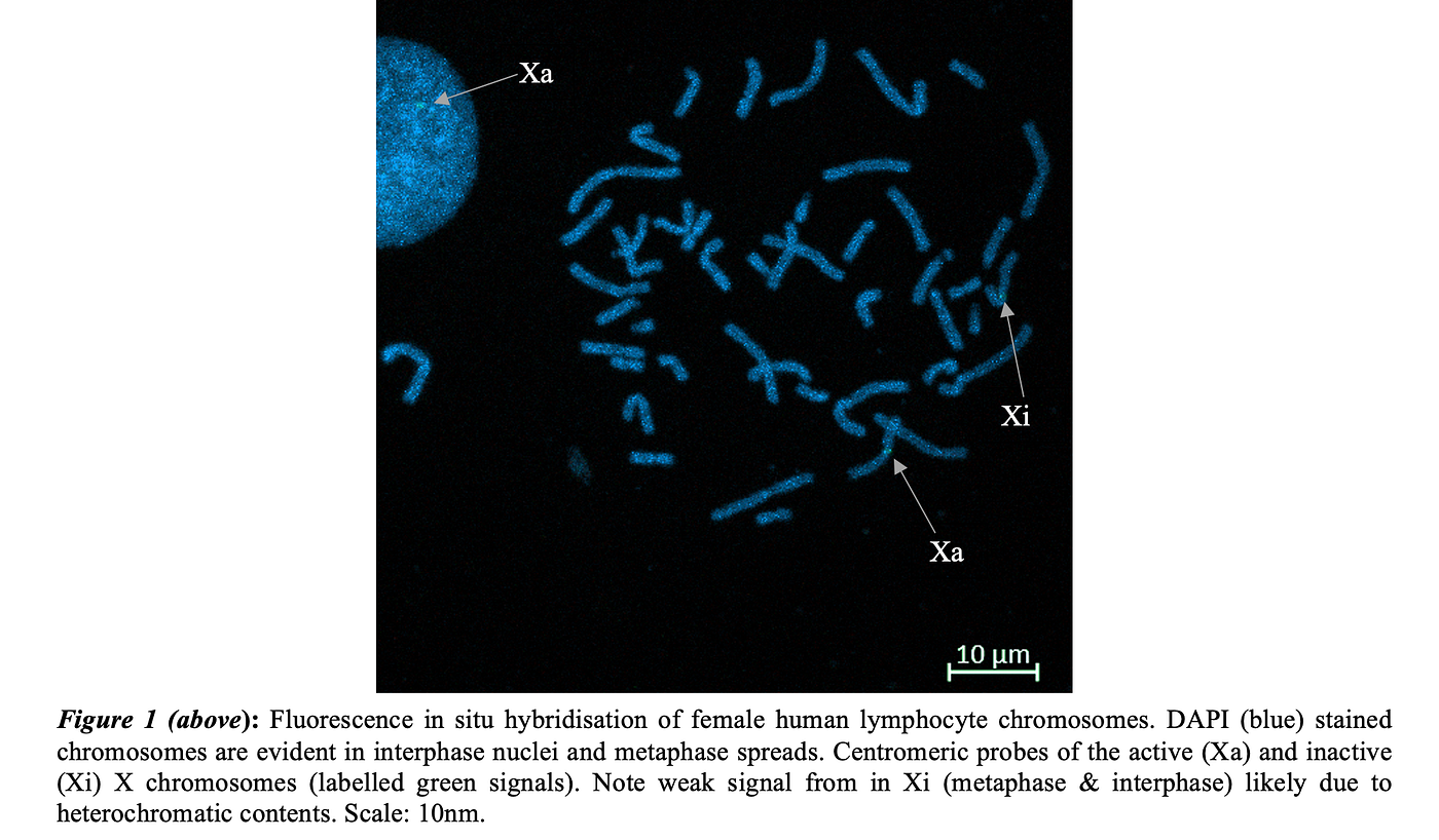


Human Fluorescence in situ Hybridisation
Human males contain sex determining chromosomes ‘X’ and ‘Y’, whilst females possess two X chromosomes. This difference represents a paradigm of epigenetic control to confirm equitable gene expression between sexes, essential for mammalian development (Rodriguez & Borensztein 2023). In females, a mechanism of ‘dosage compensation’ is performed, involving the random inactivation of one (paternal or maternal) X chromosome, which condenses to form a transcriptionally inert ‘Barr body’ (Baudat, et al, 2013). During interphase, the active X chromosome (Xa), can be observed in the nuclear interior (which contains majority of euchromatic material), whilst the inactive X (Xi) is in the nuclear periphery (mostly heterochromatic material) (Keeney, et al, 2014). However, due to technical limitations and the highly condensed (heterochromatic) nature of the Xi, there is an unreliable visualisation or ineffective binding of probes seen in figure 1, although interpretation is attempted (Cremer 2002; Mirny & Dekker 2021). The centromeric probes of the Xa chromosome can be observed (tagged green to the centromeric sequence) in the interphase and metaphase spread, due to its euchromatic contents permitting a more diffuse signal (figure 1).
Figure 2 depicts the male FISH micrographs, whereby X (green & interiorly located) and Y (red & peripherally located) gonosomes are active and visible in the interphase and metaphase spread. Due to both X and Y contributing to gene expression in the cell X inactivation does not occur (Lam and Keeney, 2014). Results in the interphase spreads had habitually weak Y signals. This could be due to factors of suboptimal probe design, denaturation inefficiency or wash stringency, but is likely justified by the Y chromosome’s constituents being enriched in repetitive sequences (satellite DNA and transposable elements) forming heterochromatin and hence the accessibility of probes (Cremer 2002; Mirny & Dekker 2021). Likewise, therefore the X is evidently located towards the nucleus’ peripheral. Whilst figure 2(A) is at an earlier stage in metaphase (signified by no cohesin removal along the chromosome arms; this ensures correct bi-orientation), figure 2(B) displays a tighter, more progressed metaphase spread (Engreitz et al. 2013). Thus, the formation of the ‘meiotic sex chromosome inactivation’ (MSCI) can be visualised (figure 2(B)). This occurs during pachytene whereby gonosomes are transiently silenced and clustered in a ‘sex body’ (Turner 2015). This is undertaken by the unsynapsed regions of both chromosomes stimulating ATR kinase to phosphorylate histone H2AX (yH2AX), followed by repressive chromatin marks accumulating (e.g, H3K9me3), condensing the chromosomes at the nuclear periphery, and hence excluding RNA polymerase II for global repression of the X/Y genes until spermatogenesis (Turner 2015).
Optimising hybridisation by increasing the denaturation temperature used (to unwind heterochromatin), using probes with higher affinity and specificity to target sequences and increasing the stringency (lower salt & higher temperature to reduce non-specific binding) have the potential to enhance the results’ signal clarity. Additionally, multiplexing the FISH with probes that target XIST RNA (coats the Xi; essential for X inactivation) would aid in Barr body identification. As well, reliability of Xi and Xa identification could be enhanced by implementing immunostaining for histone marks, in particular, H3K27me3 (heterochromatin mark; enrichment on Xi) and H3K4me3 (euchromatin mark; enrichment on Xa) (Marks et al. 2009)).
Male Meiotic Mice Immunostaining
Mice (Mus muscus) have a diploid chromosome count of 40, comprised of 38 autosomes and 2 gonosomes (either XX for female, or XY for males) (Grün and Benner, 2018). The process of meiotic cell division is split into two phases — meiosis I (reductional) and meiosis II (equational), and gives rise to genetic diversity through sexual reproduction by dividing the chromosome number in half, producing four genetically distinct haploid gametes (Grün and Benner, 2018). Prophase I (subdivided into leptotene, zygotene, pachytene, diplotene & diakensis) is the most intricate step (13/14 days of meiosis (Soh et al. 2017)), ensuring homologous recombination and valid chromosome segregation.
Leptotene involves condensation of the chromosomes into thread-like structures, in addition to the formation of axial elements along each chromosome and telomere attachment to the nuclear envelope (by telomerase) (Ewelina Bolcun-Filas & Handel 2018). Cohesin loading also ensures chromatid cohesion, whilst recombination is triggered by SPO11’s double-strand breaks (Yamada et al. 2017). Figure 3(C), depicting a stage between leptotene and zygotene, shows that staining of SC formation protein complexes can allow for the visualisation of this bead-like condensation. Zygotene involves partial chromosome synapsis, that pair lengthways forming bivalents (tetrads) and the assembly of the synaptonemal complex (SC) by SYCP1/3 proteins bridging the homologs (stabilised by cohesin) (Yamada et al. 2017). Evidence of these characteristics are shown in figure 3(C), where formation of the tetrads is occurring as the SC is attaching homolog pairs.
During the leptotene to zygotene transition, the X and Y chromosomes begin to align, but only partial synapsis is completed that does not require the pseudoautosomal region (PAR) (short regions of homology between XY chromosomes) (Kauppi et al. 2011). Contrastingly, majority of humans’ XY chromosomes are non-homologous except for PARs, meaning that PAR-dependent pairing is essential for crossover and segregation; failure of this may lead to non-disjunction and aneuploidies (Kauppi et al. 2011).
Pachytene is evident in full chromosome synapsis (complete SC), and entails crossing over between non-sister chromatids (facilitated by RAD51, DMC1), forming chiasmata (sites of crossover) warranting genetic diversity (Zickler & Kleckner 2015). This synapsis can be observed at an early stage in figure 3(A) and more completed in figure 4. A lack of homology between X and Y chromosomes justifies delayed formation of the SC and is denoted by lighter bands on the micrograph (Handel & Schimenti 2010).
Pictured in figure 3(B), the diplotene stage includes SC disassembly (thinner and less consistent red signal) (homologs remain attached at chiasmata) causing slight decondensation and separation, although cohesin complexes remain at the centromeres (shielded by shugoshin (SGO1)) maintaining sister chromatid cohesion (Clift & Marston 2011). Additionally, securin protects arm cohesion (seperase inhibition) from premature cleavage (Zickler & Kleckner 1999). Finally, diakinesis involves chromosome re-condensation in preparation for metaphase I (by condensin II), nuclear envelope breakdown and telomere release, and spindle fibre attachment to kinetochores (Chan, Severson & Meyer 2004). Whilst arms are cleaved by seperase, shugoshin protects centromeres until anaphase, to permit homolog segregation (Clift & Marston 2011).
Improvement of this study could involve multiplexing the FISH with the immunostaining protocol in either mice or humans (depending on investigation), permitting XY identification during pachytene, hence enabling SC architecture spatial mapping. This would enhance the interpretability of chromosomal and protein localisation results by simultaneously labelling DNA sequences and meiotic proteins, allowing for more accurate gonosome identification, and correlation of chromosomal structure with gene or protein expression, thus providing a more comprehensive understanding (Sorkin et al. 2025).
Conclusion
This study underscores the unique behaviour of sex chromosomes (gonosomes) in humans and mice, exhibiting how FISH and immunostaining deliver insights into organisation and meiotic dynamics that are complementary and offset limitations of one another. While FISH was able to map chromosomal location, immunostaining exposed the structural and functional aspects of prophase I. Ultimately, inferences drawn from these techniques advance out cytogenetic understanding, whilst also signifying the analytical benefit of using them in combination.
References
Baudat, F., Imai, Y. and de Massy, B., 2013. Meiotic recombination in mammals: localization and regulation. Nature Reviews Genetics, 14(11), pp.794–806. Cahoon, C.K. and Hawley, R.S. (2016). Regulating the construction and demolition of the synaptonemal complex. Nature Structural & Molecular Biology, [online] 23(5), pp.369–377. doi:https://doi.org/10.1038/nsmb.3208. Chan, R.C., Severson, A.F. and Meyer, B.J. (2004). Condensin restructures chromosomes in preparation for meiotic divisions. The Journal of Cell Biology, [online] 167(4), pp.613–625. doi:https://doi.org/10.1083/jcb.200408061. Clift, D. and Marston, A.L. (2011). The Role of Shugoshin in Meiotic Chromosome Segregation. Cytogenetic and Genome Research, [online] 133(2–4), pp.234–242. doi:https://doi.org/10.1159/000323793. Cremer, T. (2002). Chromosome territories and the functional nuclear architecture. Cytometry, [online] pp.23–23. Available at: https://www.researchgate.net/publication/295450644_Chromosome_territories_and_the_functional_nuclear_architecture [Accessed 5 May 2025]. Engreitz, J.M., Pandya-Jones, A., McDonel, P., Shishkin, A., Sirokman, K., Surka, C., Kadri, S., Xing, J., Goren, A., Lander, E.S., Plath, K. and Guttman, M. (2013). The Xist lncRNA Exploits Three-Dimensional Genome Architecture to Spread Across the X Chromosome. Science, 341(6147), pp.1237973–1237973. doi:https://doi.org/10.1126/science.1237973. Ewelina Bolcun-Filas and Handel, M.A. (2018). Meiosis: the chromosomal foundation of reproduction. Biology of Reproduction, [online] 99(1), pp.112–126. doi:https://doi.org/10.1093/biolre/ioy021. Gravholt, C.H., Andersen, N.H., Conway, G.S., Dekkers, O.M., Geffner, M.E., Klein, K.O., Lin, A.E., Mauras, N., Quigley, C.A., Rubin, K., Sandberg, D.E., Sas, T.C.J., Silberbach, M., Söderström-Anttila, V., Stochholm, K., van Alfen-van derVelden, J.A., Woelfle, J., Backeljauw, P.F. and , (2017). Clinical practice guidelines for the care of girls and women with Turner syndrome: proceedings from the 2016 Cincinnati International Turner Syndrome Meeting. European Journal of Endocrinology, [online] 177(3), pp.G1–G70. doi:https://doi.org/10.1530/eje-17-0430. Grün, F. and Benner, C., 2018. Centrioles and their role in cell division. Biochemical Society Transactions, 46(6), pp.1413–1425. Groth, K.A., Skakkebæk, A., Høst, C., Claus Højbjerg Gravholt and Anders Bojesen (2012). Klinefelter Syndrome — A Clinical Update. The Journal of Clinical Endocrinology & Metabolism, [online] 98(1), pp.20–30. doi:https://doi.org/10.1210/jc.2012-2382. Handel, M.A. and Schimenti, J.C. (2010). Genetics of mammalian meiosis: regulation, dynamics and impact on fertility. Nature Reviews Genetics, [online] 11(2), pp.124–136. doi:https://doi.org/10.1038/nrg2723. Huff, J. (2015). The Airyscan detector from ZEISS: confocal imaging with improved signal-to-noise ratio and super-resolution. Nature Methods, [online] 12(12), pp.i–ii. doi:https://doi.org/10.1038/nmeth.f.388. Hughes, J.F. and Page, D.C. (2015). The Biology and Evolution of Mammalian Y Chromosomes. Annual Review of Genetics, [online] 49(1), pp.507–527. doi:https://doi.org/10.1146/annurev-genet-112414-055311. Kauppi, L., Barchi, M., Baudat, F., Romanienko, P.J., Keeney, S. and Jasin, M. (2011). Distinct Properties of the XY Pseudoautosomal Region Crucial for Male Meiosis. Science, [online] 331(6019), pp.916–920. doi:https://doi.org/10.1126/science.1195774. Keeney, S., Lange, J. and Mohibullah, N., 2014. Self-organization of meiotic recombination initiation: general principles and molecular pathways. Annual Review of Genetics, 48, pp.187–214. Lam, I. and Keeney, S., 2014. Mechanism and regulation of meiotic recombination initiation. Cold Spring Harbor Perspectives in Biology, 7(1), p.a016634. Liehr, T. (2009). Fluorescence in situ hybridization (FISH) : application guide. Berlin ; London: Springer. Marks, H., Chow, J.C., Denissov, S., Françoijs, K.-J., Brockdorff, N., Heard, E. and Stunnenberg, H.G. (2009). High-resolution analysis of epigenetic changes associated with X inactivation. Genome Research, [online] 19(8), pp.1361–1373. doi:https://doi.org/10.1101/gr.092643.109. Mirny, L. and Dekker, J. (2021). Mechanisms of Chromosome Folding and Nuclear Organization: Their Interplay and Open Questions. Cold Spring Harbor Perspectives in Biology, [online] 14(7), p.a040147. doi:https://doi.org/10.1101/cshperspect.a040147. Soh, Y.Q.S., Mikedis, M.M., Kojima, M., Godfrey, A.K., de Rooij, D.G. and Page, D.C. (2017). Meioc maintains an extended meiotic prophase I in mice. PLOS Genetics, 13(4), p.e1006704. doi:https://doi.org/10.1371/journal.pgen.1006704. Sorkin, J., Tilton, K., Lawlor, M.A., Sarathy, S.N., Liang, S., Albanese, A., Rabbani, M., Hammoud, S.S., Ellison, C.E., Pratto, F. and Jain, D. (2025). Intercellular bridges are essential for transposon repression and meiosis in the male germline. Nature Communications, [online] 16(1). doi:https://doi.org/10.1038/s41467-025-56742-9. Turner, J.M.A. (2015). Meiotic Silencing in Mammals. Annual Review of Genetics, [online] 49(1), pp.395–412. doi:https://doi.org/10.1146/annurev-genet-112414-055145. Yamada, S., Kim, S., Tischfield, S.E., Jasin, M., Lange, J. and Keeney, S. (2017). Genomic and chromatin features shaping meiotic double-strand break formation and repair in mice. Cell Cycle, 16(20), pp.1870–1884. doi:https://doi.org/10.1080/15384101.2017.1361065. Zickler, D & Kleckner, N 1999, ‘Meiotic Chromosomes: Integrating Structure and Function’, Annual Reviews, Annual Review of Genetics, vol. 33, no. 1, pp. 603–754, viewed 5 May 2025, <https://pubmed.ncbi.nlm.nih.gov/10690419/>. Zickler, D. and Kleckner, N. (2015). Recombination, Pairing, and Synapsis of Homologs during Meiosis. Cold Spring Harbor Perspectives in Biology, [online] 7(6), p.a016626. doi:https://doi.org/10.1101/cshperspect.a016626.
메타데이터
- post_id
- 19ec6d85cbc2
- slug
- decoding-sex-chromosome-dynamics-a-dual-approach-to-cytogenetic-analyses-of-mammalian-models-using-19ec6d85cbc2
- url
- https://medium.com/@billyt13/decoding-sex-chromosome-dynamics-a-dual-approach-to-cytogenetic-analyses-of-mammalian-models-using-19ec6d85cbc2
- canonical_url
- https://medium.com/@billyt13/decoding-sex-chromosome-dynamics-a-dual-approach-to-cytogenetic-analyses-of-mammalian-models-using-19ec6d85cbc2
- author_url
- https://medium.com/@billyt13
- status
- ok
- fetched_at
- 2026-06-22 05:41:33