Beyond PRP: The Molecular Mechanics of Exosomes in Tissue Regeneration and Aesthetics
Platelet-Rich Plasma changed aesthetic medicine by turning a patient’s own blood into a therapeutic tool. Draw blood, spin it down…
Beyond PRP: The Molecular Mechanics of Exosomes in Tissue Regeneration and Aesthetics
Platelet-Rich Plasma changed aesthetic medicine by turning a patient’s own blood into a therapeutic tool. Draw blood, spin it down, concentrate the platelets, reinject — and watch degranulating platelets flood the tissue with PDGF, TGF-β, VEGF, and EGF in a rough approximation of a wound-healing cascade. For over a decade, this was as close to precision regenerative medicine as aesthetic practice had. It still works. But it is, by design, a blunt instrument: a whole biological cocktail delivered in hope that the surrounding tissue interprets it correctly.
A 3D representation of an exosome, highlighting its nanoscale structure, lipid bilayer, and carefully packaged biological payload.
Exosomes do not ask the tissue to interpret anything. They tell it exactly what to do.
This is the shift underway in regenerative dermatology right now. PRP is increasingly understood as a first-generation biologic — effective, autologous, but variable and unrefined. Exosomes represent the next iteration: not a crude tissue extract, but a curated packet of molecular instructions, engineered by nature and now harnessed by medicine, that command cellular behavior with a specificity PRP was never built to achieve.
PRP asks the body to heal itself. Exosomes hand it the instructions for exactly how.
The Anatomy of a Molecular Messenger
An exosome is not a cell, a drug, or a growth factor in the conventional sense. It is a nanoscale extracellular vesicle — a subset of the broader category of extracellular vesicles (EVs) — measuring roughly 30 to 150 nanometers in diameter, small enough that dozens could fit inside a single red blood cell. Each exosome is enclosed by a lipid bilayer membrane, giving it the structural integrity to survive transit through the extracellular space and the biophysical machinery to fuse with, or be internalized by, a target cell.

Exosome biogenesis: The endosomal pathway illustrates how multivesicular bodies (MVBs) fuse with the plasma membrane to release targeted exosomes into the extracellular space.
Their origin is intracellular. Exosomes are generated through the endosomal pathway: the membrane of a maturing endosome buds inward on itself, producing intraluminal vesicles inside a structure known as a multivesicular body (MVB). When the MVB fuses with the plasma membrane, those intraluminal vesicles are released into the extracellular environment as exosomes — no longer cellular debris, but deliberately packaged cargo. As Kalluri and LeBleu detailed in their landmark 2020 review in Science, this biogenesis pathway allows exosomes to carry a payload that faithfully reflects the biological state and intent of the cell that produced them.
That payload is where the therapeutic power lives. Inside the vesicle, protected from extracellular degradation, exosomes carry:
- Messenger RNA (mRNA) — transcripts that can be translated into functional protein once inside a recipient cell
- MicroRNA (miRNA) — short non-coding RNA sequences that post-transcriptionally silence or modulate specific target genes
- Cytokines — signaling proteins that regulate inflammation, immune activity, and cell-to-cell communication
- Growth factors — including IGF-1, HGF, KGF, VEGF, and TGF-β, which drive proliferation, angiogenesis, and matrix synthesis
Surface markers such as CD9, CD63, CD81, and TSG101 identify the vesicle as a bona fide exosome and mediate its docking onto target cell membranes — the molecular handshake that precedes cargo delivery.
Paracrine Signaling: Command Without Colonization
The therapeutic logic of exosomes rests on a concept called paracrine signaling — the process by which a cell influences the behavior of nearby cells through secreted molecules, without needing to physically integrate into the target tissue itself. This is the mechanism that made mesenchymal stem cell (MSC) therapy compelling in the first place: it was never really the transplanted stem cells doing the regenerative work by differentiating and replacing damaged tissue. It was what they secreted.

Paracrine signaling pathways demonstrating how stem cell-derived exosomes and soluble factors actively modulate surrounding tissue and immune responses without cellular integration.
Exosomes isolate that secretory function and deliver it directly, cell-free.
Once released, an exosome travels through the extracellular matrix and is taken up by a recipient cell — via direct membrane fusion, receptor-mediated endocytosis, or macropinocytosis. Upon internalization, its cargo is released into the recipient cell’s cytoplasm, where miRNA can suppress specific mRNA transcripts, delivered mRNA can be translated into new protein, and growth factors can trigger downstream signaling cascades. The recipient cell’s gene expression and protein synthesis shift in response — not because a new cell has arrived to do the work, but because an existing, often aged or senescent, cell has received new instructions.
The therapeutic agent never needs to survive engraftment, evade immune clearance, or persist in the tissue. Only its message does.
This is the conceptual leap beyond PRP: PRP delivers growth factors that transiently bathe the tissue in a general pro-healing signal. Exosomes deliver a specific genetic and proteomic instruction set that reprograms the target cell’s behavior at the transcriptional level — a fundamentally more precise mode of intervention.
Exosomes vs. PRP: Why Precision Outperforms Volume
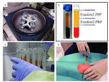
PRP’s central limitation is structural: it is only ever as good as the blood it comes from. The concentration, quality, and inflammatory profile of a patient’s platelets are downstream of variables the clinician cannot control — chronological age, systemic inflammation, medication use, metabolic health, and hormonal status all shift the composition of what gets reinjected. A PRP preparation drawn from a 60-year-old with subclinical inflammatory burden is not biologically equivalent to one drawn from a 30-year-old, even when the injection technique is identical. PRP can also carry pro-inflammatory cytokines alongside its regenerative growth factors, particularly in patients with underlying metabolic or autoimmune conditions — an inherent, uncontrollable variable in an autologous product.

Traditional Platelet-Rich Plasma (PRP) preparation relies on isolating fractions of the patient’s own blood, meaning its regenerative potential is limited by the biological baseline of the donor.
Exosomes sidestep this problem by decoupling the therapeutic agent from the patient’s own biology entirely. Most clinical-grade exosome products are derived from carefully screened, allogeneic mesenchymal stem cell lines — commonly adipose-derived or umbilical cord-derived — cultured under controlled conditions and isolated via ultracentrifugation or size-exclusion chromatography. The result is a product that can be:
- Standardized — quantified by particle concentration and characterized by surface marker profile, batch to batch
- Highly concentrated — delivering a dose of regenerative signaling molecules far beyond what a single patient’s own plasma could yield
- Purely regenerative — free of the inflammatory variability and cellular debris that autologous blood products inevitably carry
- Cell-free — eliminating concerns around cellular immune rejection or tumorigenic potential associated with live-cell therapies

Split-face clinical trials offer direct comparisons, revealing that standardized exosome treatments can match or exceed the efficacy of standard modalities without the variable inflammatory profile.
This is no longer theoretical. In 2025, Estupiñan, Ly, and Goldberg published the first investigator-blinded, split-face trial directly comparing adipose MSC-derived exosomes against PRP for photoaged facial skin, using radiofrequency microneedling as the delivery vehicle on each side of the face. The result was a non-inferiority finding: both modalities produced comparable improvements in wrinkling, dyschromia, erythema, and texture, with histological confirmation of increased collagen I and glycosaminoglycan content on both sides — no significant difference between arms. The clinical implication is notable less for exosomes “beating” PRP than for what it removes from the equation: no phlebotomy, no centrifugation, and a standardized product unaffected by the patient’s own blood quality on the day of treatment.
Clinical Mechanics I: Skin Rejuvenation at the Fibroblast Level
The dermal fibroblast is the effective target of nearly every skin rejuvenation strategy, and exosome signaling reaches it with unusual specificity. Upon uptake, exosomal cargo activates intracellular signaling pathways that increase transcription of COL1A1 and COL3A1, driving synthesis of type I and type III collagen, while simultaneously upregulating elastin and fibrillin expression to rebuild the elastic fiber network responsible for skin recoil. In parallel, exosome-delivered signals suppress matrix metalloproteinases — particularly MMP-1 and MMP-9 — the enzymes chiefly responsible for degrading collagen in photoaged and chronologically aged skin.

The structural contrast between young and aged skin, highlighting the accumulation of senescent cells that exosomes actively target.
The second, less obvious mechanism concerns cellular senescence. As dermal fibroblasts age, a subset enters a senescent state, ceasing proliferation while adopting a senescence-associated secretory phenotype that floods the surrounding matrix with inflammatory mediators and further degrades tissue architecture — a self-perpetuating cycle of dermal decline. In their 2024 review in Cell Communication and Signaling, Hajialiasgary Najafabadi and colleagues detail how stem cell-derived exosomes intervene directly in this cycle: decreasing MMP expression, increasing collagen and elastin output, and modulating the intracellular signaling pathways associated with oxidative stress and senescence itself. The effect is not merely stimulatory. It is corrective — nudging an aged, secretory-dysfunctional fibroblast population back toward a proliferative, matrix-synthesizing phenotype.

Exosomes directly target dermal fibroblasts, upregulating the genes and ribosomal machinery responsible for synthesizing high volumes of structural collagen fibers.
Clinical Mechanics II: Hair Restoration and the Anagen Switch
The biology of hair loss is fundamentally a biology of cycling failure. Follicles do not die in androgenetic alopecia so much as they get trapped — shorter and shorter periods in anagen (the active growth phase), longer periods in telogen (the resting phase), progressively miniaturizing with each cycle. The dermal papilla cell (DPC), sitting at the base of the follicle, governs this cycling through its own paracrine output — and DPC-derived exosomes appear to be the vehicle for that governance.

The human hair growth cycle. Exosomal therapy aims to reset cycling behavior, rescuing miniaturized follicles from prolonged telogen and extending the active anagen phase.
In a 2018 study published in Biochemical and Biophysical Research Communications, Zhou and colleagues isolated exosomes from cultured dermal papilla cells — approximately 105 nanometers in diameter and positive for the exosomal markers CD9, CD63, and TSG101 — and injected them cutaneously into mouse follicles at different points in the hair cycle. The result was a measurable acceleration of the transition into anagen and a delay in the onset of catagen (the regression phase), an effect the authors traced to activation of β-catenin and Sonic hedgehog signaling in outer root sheath cells.
A separate 2019 study in Experimental Dermatology by Kwack and colleagues extended this further using exosomes derived from three-dimensionally cultured dermal papilla spheres. These exosomes increased proliferation of both dermal papilla and outer root sheath cells, elevated expression of IGF-1, KGF, and HGF within DP cells, and — critically — increased hair shaft elongation in cultured human hair follicles ex vivo. Local injection in mice induced anagen directly from telogen and prolonged the anagen phase beyond baseline duration.
Together, these findings describe a therapy that does not simply nourish an existing follicle, but actively resets its cycling behavior — waking dormant follicles trapped in prolonged telogen and extending the growth window once anagen is reached. It is the same underlying principle at work in skin: a dormant or dysfunctional cell population, receiving a precise molecular signal, resumes a younger functional program without the need for any new cell to take up residence.

The future of regenerative aesthetics lies in omic profiling and biological precision, utilizing cell-free messengers to rewrite cellular behavior with unprecedented accuracy.
Scientific References
- Kalluri R, LeBleu VS. The biology, function, and biomedical applications of exosomes. Science. 2020;367(6478):eaau6977. doi:10.1126/science.aau6977. PMID: 32029601.
- Zhou L, Wang H, Jing J, Yu L, Wu X, Lu Z. Regulation of hair follicle development by exosomes derived from dermal papilla cells. Biochemical and Biophysical Research Communications. 2018;500(2):325–332. doi:10.1016/j.bbrc.2018.04.067. PMID: 29654758.
- Kwack MH, Seo CH, Gangadaran P, Ahn BC, Kim MK, Kim JC, Sung YK. Exosomes derived from human dermal papilla cells promote hair growth in cultured human hair follicles and augment the hair-inductive capacity of cultured dermal papilla spheres. Experimental Dermatology. 2019;28(7):854–857. doi:10.1111/exd.13927. PMID: 30924959.
- Hajialiasgary Najafabadi A, Soheilifar MH, Masoudi-Khoram N. Exosomes in skin photoaging: biological functions and therapeutic opportunity. Cell Communication and Signaling. 2024;22(1):32. doi:10.1186/s12964–023–01451–3. PMID: 38217034.
- Estupiñan B, Ly K, Goldberg DJ. Adipose mesenchymal stem cell-derived exosomes versus platelet-rich plasma treatment for photoaged facial skin: an investigator-blinded, split-face, non-inferiority trial. Journal of Cosmetic Dermatology. 2025. doi:10.1111/jocd.70208. PMID:40414798
메타데이터
- post_id
- 1bc6e9e6acbf
- slug
- beyond-prp-the-molecular-mechanics-of-exosomes-in-tissue-regeneration-and-aesthetics-1bc6e9e6acbf
- url
- https://medium.com/@dr.jannat.tech/beyond-prp-the-molecular-mechanics-of-exosomes-in-tissue-regeneration-and-aesthetics-1bc6e9e6acbf
- canonical_url
- https://medium.com/@dr.jannat.tech/beyond-prp-the-molecular-mechanics-of-exosomes-in-tissue-regeneration-and-aesthetics-1bc6e9e6acbf
- author_url
- https://medium.com/@dr.jannat.tech
- status
- ok
- fetched_at
- 2026-07-11 03:42:06