Brief Review — Automatic Left Ventricular Outflow Tract Classification for Accurate Cardiac MR…
5-Layer CNN for Left Ventricular Outflow Tract (LVOT) Classification
Brief Review — Automatic Left Ventricular Outflow Tract Classification for Accurate Cardiac MR Planning
5-Layer CNN for Left Ventricular Outflow Tract (LVOT) Classification
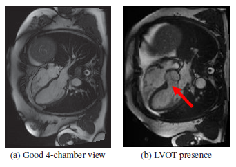
Presence of LVOT Causes Inaccurate Cardiac MR Planning
**Automatic Left Ventricular Outflow Tract Classification for Accurate Cardiac MR Planning 5-Layer CNN, by King’s College London, Hospital NHS Foundation Trust, and Imperial College London 2018 ISBI **(Sik-Ho Tsang @ Medium)
Quality Assessment 2020 [Mean Teacher + ROI Consistency] 2021 [Model Fusion + SVR] 2022 [Swin-MIQA] ==== My Healthcare and Medical Related Paper Readings ==== ==== My Other Paper Readings Are Also Over Here ====
- One result of inaccurate cardiac MR planning is an ‘off-axis’ orientation of the 4-chamber view, often recognized by the presence of the left ventricular outflow tract (LVOT).
- A 5-layer CNN is proposed to automatically detect the presence of the LVOT in cardiac MRI.
Outline
- 5-Layer CNN
- Results
1. 5-Layer CNN
- The proposed framework of using a CNN for LVOT detection is as follows
- Contrast normalization of the target images: Given a full CMR image we first normalize the pixel values between 0 and 1.
- Image cropping using atrial masks: A template-matching based region of interest (ROI) extraction is used to crop a 64×64 block. 100 MR images are independent from LVOT detection algorithm validation dataset, are solely used for ROI extraction.
- CNN image classification of correct versus off-axis planning of the 4-chamber images: A CNN of 64×64 — 60×60×32 — 26×26×64–128–2–1 structure, is used.
- The first layer is a convolutional layer, which filters the input with 32 kernels, each of size 5×5. The convolutional layer produces 32 feature maps, each of size 60×60, followed by a 2×2 pooling operation that reduces each feature map to one max.
- Two fully connected (dense) layers (one with 128 and one with 2 nodes) come after the pooling.
- The last layer is a simple softmax classification layer with a one dimensional output that predicts the class.
- **ReLU** is used.
- **Dropout **with a probability of 0.5 at all convolutional layers and after the first fully connected layer to enforce regularization.
- Data augmentation of random rotation and translation is used.
2. Results
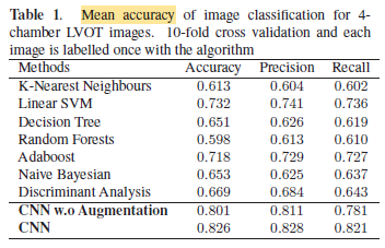
Mean accuracy of image classification for 4-chamber LVOT images.
- 82.6% accuracy is obtained by the proposed CNN with data augmentation.
메타데이터
- post_id
- 4e1f9e873ee2
- slug
- brief-review-automatic-left-ventricular-outflow-tract-classification-for-accurate-cardiac-mr-4e1f9e873ee2
- url
- https://medium.com/@sh-tsang/brief-review-automatic-left-ventricular-outflow-tract-classification-for-accurate-cardiac-mr-4e1f9e873ee2
- canonical_url
- https://medium.com/@sh-tsang/brief-review-automatic-left-ventricular-outflow-tract-classification-for-accurate-cardiac-mr-4e1f9e873ee2
- author_url
- https://medium.com/@sh-tsang
- status
- ok
- fetched_at
- 2026-06-29 01:02:39