Image Processing, Segmentation, Classification, and Localization of Medical Image-3
This is the continuation of this Medical Images task series: [Link]
Image Processing, Segmentation, Classification, and Localization of Medical Image-3
This is the continuation of this Medical Images task series: [Link]
Task 6: Segmentation 2- Lung area extraction from chest X-ray images (lung area: 255/heart: 85/outside lung:170/outside the body: 0)
The objective of this project was to develop a lung area segmentation model for chest X-ray images. The labels in the dataset contained pixel values representing differ- ent anatomical regions, including the lung area (255), heart (85), outside the lung (170), and outside the body (0). This report provides a comprehensive overview of the code im- plementation and its results.
6.1. Model Architecture:
The code implemented a custom Fully Convolutional Network (FCN) model designed for semantic segmentation tasks. This choice of architecture, while straightforward, may have limitations in capturing intricate image details.
6.2. Custom Dataset Handling:
A custom dataset class, CustomDataset, was developed to load and preprocess both chest X-ray images and their corresponding label images. The labels were preprocessed to map pixel values to specific anatomical classes (heart, outside lung, lung, and outside the body).
6.3. Training and Optimization:
The model underwent training for 30 epochs with a batch size of 16, using the Adam optimizer with a learning rate of 0.001. Cross-entropy loss was employed as the optimiza- tion criterion, a common choice for multi-class segmentation tasks.
6.4. Training Progress:
An observation in the code is the repetition of the train- ing loop three times, which may lead to redundant computations and resource consumption.
Here is the sample normal label and color coded Image:

6.5. Suggestions for Improvement:
Model Architecture: To enhance model performance, consider exploring more complex network architectures, such as deeper FCN variants or established semantic segmentation models like U-Net. These architectures are better equipped to capture fine-grained image details.
Hyper parameter Tuning: Fine-tuning hyper parameters like the learning rate and batch size can improve model convergence and generalization. Careful selection of these values can lead to better results.
Evaluation Metrics: Integrating evaluation metrics like Intersection over Union (IoU) or Dice coefficient would provide a more comprehensive assessment of segmentation performance and facilitate model comparison.
Training Loop Optimization: Addressing the repeti- tion of the training loop in the code is essential to ensure computational efficiency and avoid unnecessary computations.
The approach presented a foundational framework for lung area segmentation in chest X-ray images. However, there is substantial room for improvement in terms of ac- curacy and precision. Enhancements to the model architecture, hyper parameter tuning, incorporation of evaluation metrics, and optimization of the training process are crucial for achieving accurate identification of anatomical regions in medical images.
Task 7: Object Localization Approaches in Chest X-ray Images
This approach shows the task of object localization within chest X-ray images. Object localization involves identifying and drawing bounding boxes around specific anatomical areas within the images. Two different approaches have been presented: one using color-based seg- mentation and another employing a YOLO (You Only Look Once) deep learning model for object detection.
7.1. Color-Based Segmentation:
This approach utilizes color-based segmentation to iden- tify and localize anatomical areas within chest X-ray im- ages.It processes a directory of label images where each pixel’s color corresponds to specific anatomical regions: lung area (white), heart area (gray), outside the lung (light gray), and outside the body (black).Bounding boxes are drawn around each region to visually highlight their loca- tions.
7.2. Usage:
The method reads label images, processes pixel colors, and identifies regions based on predefined color values. It employs OpenCV for image processing and contour detection to create bounding boxes. The results are displayed using the cv2imshow function in a Colab environment.

7.3. YOLO-Based Object Detection:
The second approach utilizes a pre-trained YOLOv3 (You Only Look Once version 3) model for object detec- tion and localization in chest X-ray images.YOLOv3 is a popular deep learning model for real-time object detection. It loads a custom-trained YOLOv3 model and uses it to de- tect and localize objects from a given image. The method is specifically tailored to identify and localize objects corre- sponding to classes such as ”lung,” ”heart,” ”outside lung,” and ”outside body.”
7.4. Usage:
The code loads a pre-trained YOLOv3 model along with class labels and colors.It processes the input image, per- forms object detection, and draws bounding boxes around detected objects. Detected objects are labeled with their class names and confidence scores. The resulting image with bounding boxes is displayed using Matplotlib.
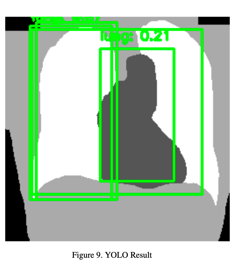
The YOLO-based object detection approach offers the advantage of utilizing a deep learning model trained to rec- ognize and localize objects. However, the provided code may have limitations in achieving satisfactory localization results.
7.5. Areas of Improvement:
Fine-Tuning for Medical Images: Considering fine- tuning the YOLOv3 model on a dataset of chest X-ray images to adapt it to the specific characteristics of medical imaging. Fine-tuning can enhance the model’s ability to localize anatomical regions accurately.
Threshold Optimization: Experimenting with different confidence thresholds to find the optimal value that balances precision and recall. This adjustment can help improve localization accuracy.
Evaluation Metrics: Incorporating evaluation metrics such as Intersection over Union (IoU) or Dice coefficient to quantitatively assess the quality of object localization. These metrics can provide insights into the model’s performance and guide improvements.
In conclusion, while both color-based segmentation and YOLO-based object detection offer valuable approaches for object localization in chest X-ray images, there is room for improvement to achieve more accurate and clinically meaningful results. Addressing the mentioned areas of improvement can lead to enhanced localization performance, making these techniques more valuable in medical image analysis.
Conclusion
In summary, By integrating both image loading and dis- play tasks, this combined task demonstrates the ability to handle diverse medical image formats. Our image classification model successfully predicts the direction of chest X- ray images with a high level of accuracy. The combination of data augmentation, dropout layers, and other techniques contributed to the model’s ability to generalize well to un- seen data. Future work may involve exploring more advanced architectures, optimizing hyperparameters, and expanding the dataset to further enhance the model’s perfor- mance and generalization capabilities.
Furthermore, our gender classification model accurately distinguishes between male and female patients based on chest X-ray images, a vital step for patient profiling and personalized healthcare. Additionally, our age estimation model demonstrates the potential to predict patient ages accurately, contributing to diagnosis and treatment planning. Moreover, our segmentation reports in Task 5 and Task 6 have illuminated the versatility of image processing techniques in medical image analysis. While Task 5 focused on lung area extraction, Task 6 extended the segmentation to four distinct areas. These methods have provided valuable insights, and future work may concentrate on enhancing ro- bustness and achieving pixel-level accuracy in localization.
Additionally, in Task 7, we employed YOLO-based ob- ject detection for localization within chest X-ray images. While YOLOv3 is a powerful model, it may benefit from fine-tuning on medical image data to enhance localization accuracy. Adjusting confidence thresholds and incorporat- ing evaluation metrics can further improve the localization results
As we look ahead, there is room for continued advancement in the field of medical image analysis. Future work may encompass the exploration of more advanced model architectures, fine-tuning of hyperparameters, and the expansion of the dataset. These endeavors aim to further en- hance the model’s performance and generalization capabilities, ultimately advancing the state-of-the-art in medical im- age analysis.
In conclusion, this research lays a solid foundation for innovative image analysis techniques in healthcare.
메타데이터
- post_id
- 58992b893bfe
- slug
- image-processing-segmentation-classification-and-localization-of-medical-image-3-58992b893bfe
- url
- https://medium.com/@vignesh.g1609/image-processing-segmentation-classification-and-localization-of-medical-image-3-58992b893bfe
- canonical_url
- https://medium.com/@vignesh.g1609/image-processing-segmentation-classification-and-localization-of-medical-image-3-58992b893bfe
- author_url
- https://medium.com/@vignesh.g1609
- status
- ok
- fetched_at
- 2026-06-15 20:49:13