Spatial and Single-Cell Transcriptomics Reveal a Cancer-Associated Fibroblast Subset in HNSCC That…
Research Article Digest
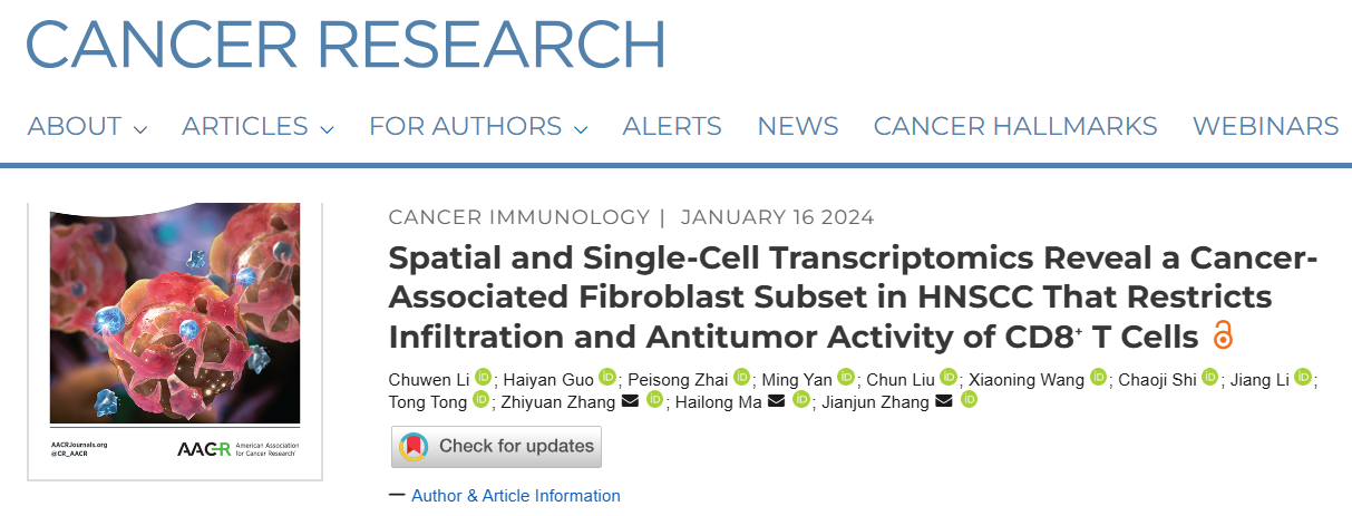
Spatial and Single-Cell Transcriptomics Reveal a Cancer-Associated Fibroblast Subset in HNSCC That Restricts Infiltration and Antitumor Activity of CD8+ T Cells
Research Article Digest
Journal: Cancer Research
Affiliations: Zhang et. al., School of Medicine, Shanghai Jiao Tong University, Shanghai
Summary
This research article examines specific cancer-associated fibroblast (CAF) subpopulations associated with limited CD8+ T cell infiltration and antitumor activity in head and neck squamous cell carcinoma (HNSCC).
Role of CD8+ T Cells in HNSCC
- Immunotherapy, while promising for cancer treatment, has limitations in treating HNSCC due to low response rates. The infiltration of cytotoxic CD8+ T cells into tumor areas is crucial for its effectiveness, highlighting the importance of understanding the complex tumor microenvironment (TME) networks that regulate these cells’ infiltration and function.
- Cancer-associated fibroblasts (CAFs) are key components of the TME, playing multifaceted roles in regulating tumor immune responses. However, CAF heterogeneity poses challenges to strategies aimed at improving immunotherapy by modulating these cells.
Spatial Analysis Reveals CAF Subsets Associated with CD8+ T Cell Infiltration
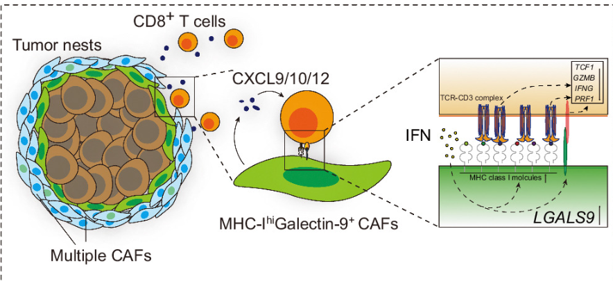
- Spatial transcriptomics analysis of HNSCC specimens with varying immune infiltration patterns identified specific CAF subsets associated with restricted CD8+ T cell infiltration. These CAFs express high levels of CXCL9, CXCL10, and CXCL12, chemokines known to attract CD8+ T cells.
- Specific expression patterns of ECM components in stromal cells associated with limited CD8+ T cell infiltration were also observed.
Single-Cell Analysis Reveals MHC-IhiGal9+ CAFs Associated with CD8+ T Cell Dysfunction
- Single-cell RNA sequencing (scRNA-seq) of five pairs of tumor and adjacent tissues examined specific CAF subsets associated with restricted CD8+ T cell infiltration and dysfunction. Results indicated high expression of MHC-I molecules (HLA-A/B/C and B2M) and galectin-9 (Gal9) in these CAFs.
- Notably, the proportion of MHC-IhiGal9+ CAFs negatively correlated with the abundance of TCF1+GZMK+ CD8+ T cell subsets, an effector T cell subpopulation.
- Findings suggest that Gal9 on CAFs induces CD8+ T cell dysfunction and reduces the proportion of tumor-infiltrating TCF1+CD8+ T cells. Additionally, IFN signaling pathways, particularly IFNβ and IFNγ, were found to upregulate galectin-9 and MHC-I molecule expression in CAFs, respectively.
Study Significance
- The study identifies the role of MHC-IhiGal9+ CAFs in immune evasion in HNSCC, potentially paving the way for developing more effective immunotherapy strategies.
- These findings suggest that targeting MHC-IhiGal9+ CAFs or Gal9 may be a promising approach to enhance antitumor immune responses in HNSCC patients.
- However, further research is needed to fully elucidate the clinical implications of these findings and explore therapeutic strategies based on modulating these CAF subsets to enhance immunotherapy.
Experimental Flow
This study investigates the impact of specific cancer-associated fibroblast (CAF) subpopulations on CD8+ T cell infiltration and function in head and neck squamous cell carcinoma (HNSCC). The research employs a multi-faceted approach:
- Bioinformatics Analysis:
- Analyzed TCGA data for CD8+ T cell infiltration and MHC-I expression in HNSCC.
- Explored correlations between key genes using TIMER and GEPIA databases.
2. Tissue Analysis:
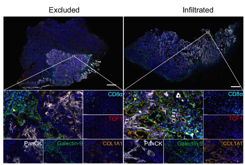
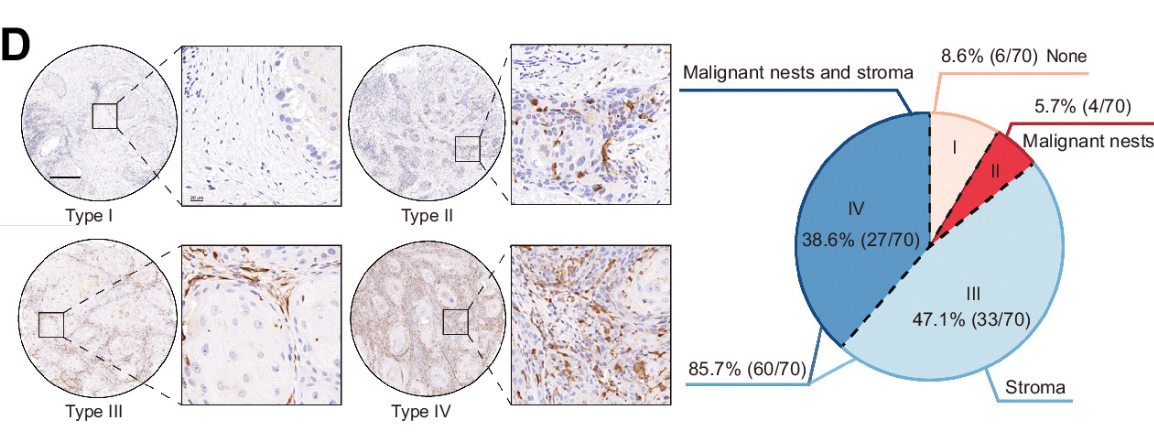
- Performed immunohistochemistry and multiplex immunofluorescence staining to examine spatial relationships between CAFs and CD8+ T cells.
- Conducted spatial transcriptomics to analyze gene expression patterns in different HNSCC immune subtypes.
3. Single-cell RNA Sequencing:
- Analyzed CAF heterogeneity and defined subpopulations.
- Examined CD8+ T cell subsets and their interactions with CAFs.
4. In Vitro Experiments:
- Performed gene knockdowns (B2M, LGALS9) in CAFs and fibroblasts.
- Conducted co-culture experiments with tumor cells and CD8+ T cells.
5. Animal Studies:
- Established subcutaneous tumor models to investigate the effects of CAF modifications on tumor growth and CD8+ T cell function.
6. Molecular Analyses:
- Used flow cytometry, real-time PCR, and Western blotting to examine protein and gene expression changes in response to various treatments and conditions.
This comprehensive approach provides insights into the role of MHC-IhiGal9+ CAFs in suppressing CD8+ T cell function in HNSCC, offering potential avenues for new immunotherapy strategies.
TissueGnostics Solutions in this Article
- TG TSA Multiplex IHC Assay Kits: Used for multiplex immunofluorescence staining(8-colored panel) of HNSCC tissue sections. This staining technique allows simultaneous detection of multiple target proteins, such as CD3, α-SMA, pan-cytokeratin (PanCK), galectin-9, CD8α, TCF1, and COL1A1 used in this study.
- TissueFAXS Spectra Systems: Employed to scan and visualize tissue sections stained with TSA kits, generating high-resolution multiplex immunofluorescence images.
- StrataQuest analysis software: Utilized for quantitative analysis of images acquired by TissueFAXS, assessing spatial relationships between different cell types, such as the distance between Gal9+ CAFs and TCF1+CD8+ T cells.

By integrating these technologies, researchers were able to:
- Confirm the spatial proximity between Gal9+ CAFs and TCF1+CD8+ T cells, suggesting that Gal9+ CAFs may influence TCF1+CD8+ T cell function through direct cell-to-cell contact.
- Quantify differences in spatial distribution of Gal9+ CAFs and TCF1+CD8+ T cells across various HNSCC immune subtypes, providing insights into the role of Gal9+ CAFs in different immune microenvironments.
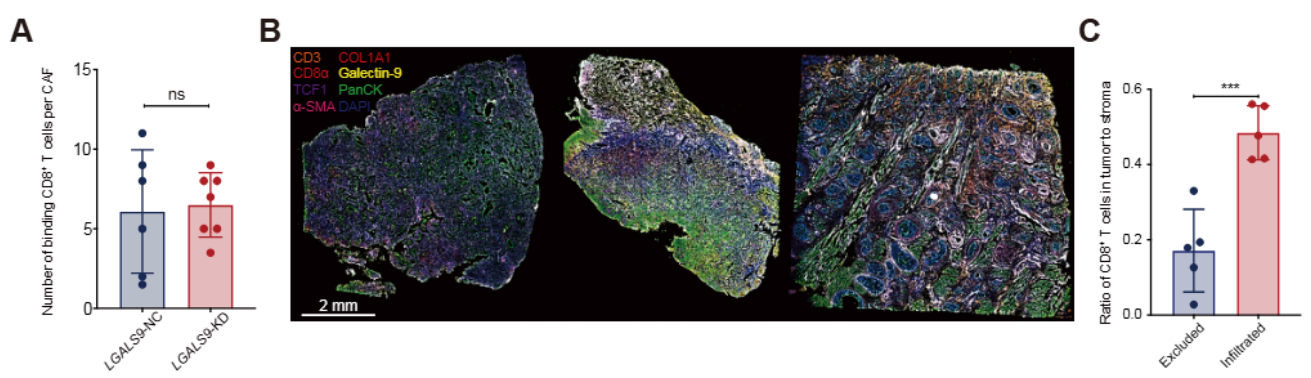
TissueGnostics products played a crucial role in this research. Their multiplex immunofluorescence staining, image scanning, and quantitative analysis capabilities helped researchers uncover the spatial relationships between Gal9+ CAFs and TCF1+CD8+ T cells, providing vital information for understanding immune evasion mechanisms in HNSCC.
Contact Us
office@tissuegnostics.cn (APAC office) or
felix.tsai@tissuegnostics.cn (Felix Tsai, APAC regional business manager)
TissueGnostics GmbH
Address: Taborstrasse 10/2/8, A-1020 Vienna, AUSTRIA, EU
Website: www.tissuegnostics.com
메타데이터
- post_id
- 645009926a4d
- slug
- spatial-and-single-cell-transcriptomics-reveal-a-cancer-associated-fibroblast-subset-in-hnscc-that-645009926a4d
- url
- https://medium.com/@TGAP_Blog/spatial-and-single-cell-transcriptomics-reveal-a-cancer-associated-fibroblast-subset-in-hnscc-that-645009926a4d
- canonical_url
- https://medium.com/@TGAP_Blog/spatial-and-single-cell-transcriptomics-reveal-a-cancer-associated-fibroblast-subset-in-hnscc-that-645009926a4d
- author_url
- https://medium.com/@TGAP_Blog
- status
- ok
- fetched_at
- 2026-07-22 16:46:20