ResNet18 in Medical Imaging: Why Deep Residual Networks See What We Miss
In the last decade, artificial intelligence (AI) in medical imaging has transitioned from pattern recognition to high fidelity…
ResNet18 in Medical Imaging: Why Deep Residual Networks See What We Miss
In the last decade, artificial intelligence (AI) in medical imaging has transitioned from pattern recognition to high fidelity visualization. Central to this change has been one simple, yet powerful architecture — ResNet18. When radiologists or pathologists view a scan with many details, they depend on years of training to separate the signal from the noise. ResNet18 is simply mimicking this process on silicon. Below, we propose reasons why ResNet18 continues to be relevant in medical image analysis, especially when precision and interpretability matter.
Why ResNet18 for Medical Imaging?
Unlike its larger, bulkier counterparts — which are burdened with needing terabytes of images to train and develop — ResNet18 has a comfort zone — small, fast and weirdly accurate.
Medical datasets like histopathology slides, CT scans, or imaging blood cells, are significantly affected by noise and changes in stains, light, and resolution.The design of ResNet18 helps reduce this impact as it learns from reusing features and residual learning, making it more resistant to noise and subtle changes in the images.
Simply put: ResNet18 never forgets. It learns and builds on what it has learned.
What is ResNet18?
ResNet refers to a Residual Network created by Microsoft Research in 2015. The “18” part indicates that it is 18 layers deep, meaning that when the model is complete, there will be 18 layers of learnable parameters.
The main idea behind ResNet is the “skip connection,” which is a smart way of allowing the model to skip layers so that the model doesn’t lose information. Instead of just stacking up layers mindlessly, ResNet learns residuals, which again signifies small meaningful differences that improve the model performance.
This addresses a fundamental issue called vanishing gradients: an issue that locks up deeper networks from learning without disintegrating
How ResNet18 Thinks: A Simple Flow
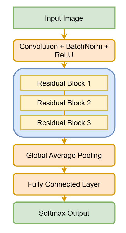
Flow of the ResNet18 architecture showing how residual blocks extract progressively complex medical imaging features — from edges to tissues to tumors.
Each block learns to detect progressively complex patterns — from edges to textures, from tissues to tumors.
What Datasets Does ResNet18 Work Well With?
ResNet18 has been been tested extensively on medical imaging datasets. For example:
- PathMNIST — colon tissue histology.
- Blood Cell Dataset — normal vs infected blood cells.
- Chest X-ray — detection of pneumonia, COVID, or other lung diseases.
These datasets often have variability due to outliers: the smallest little stain, odd texture, or slightly dim border can change a diagnosis entirely.
ResNet18’s residual design works to lessen the sensitivity to these outliers, allowing the model to learn what is “invariant” across specimens and not just what is visually obvious.
What Makes ResNet18 Powerful for Medical Imaging
-
Lightweight yet accurate, this model is the perfect fit for use in medical labs and for embedded devices performing diagnostics in restricted hardware conditions.
-
Stable training: Skip connections prevent gradient vanishing, ensuring consistent learning even on small datasets.
-
Feature reuse is highlighted — it figures out what is important (e.g. tissue boundaries, nuclei density and lesion shape) and disregards incidents of redundant noise.
-
Explainable — used in concert with visualization technologies (such as Grad-CAM and Integrated Gradients) this will easily fall into those explanations.
Thus this combination of efficiency and logic are acceptable architectures for your healthcare Ai teams.
Real-World Use Case: Blood Cell Visualization with ResNet18 + Grad-CAM
Let’s look at a practical case — Blood Cell Classification. Using ResNet18 trained on the Blood Cell Dataset, we can distinguish types of cells (eosinophil, monocyte, lymphocyte, neutrophil).

To understand why the explanations of the decision, we can use Grad-CAM (Gradient-weighted Class Activation Mapping) — a visualization technique where we show the area of the images that influenced the model to make its prediction.

Hence you will provide to the ai team three outputs:
- The Original Image — the raw input to the data set.
- The Heatmap — the areas of attentiveness that the model thought were interesting.
- The Overlay — How both image provided conclusions to decisioning.
This type visual transparency can go a long way toward building trust ans sense of accountability for ai in the clinic.
Why Pixel Contribution Matters
Each pixel in a medical image provides a part of the context. For example, in a blood smear, one pixel might represent a cell nucleus or an outline that goes from healthy to abnormal cells.
ResNet18, along with interpretability methods, allows for pixel-level quantification of importance.
- High-contribution pixels = helpful observations for diagnosis.
- Low-contribution pixels = non-diagnostic background.
However, understanding the contribution of such pixels allows the model to not only function as a black box, but to become a supportive tool in the diagnosis. The physician would be able to related a machine’s attention to medical thinking — ultimately shifting the AI role from a guesser to a collaborator.
Final Thoughts
Although ResNet18 may be close to a decade old, it is also a classic in medical imaging. Its blend of simplicity, stability, and interpretability has allowed it to remain a trusted framework for healthcare artificial intelligence systems.
From pathology slides to blood cell microscopy, ResNet18 has successfully demonstrated, sometimes clearly beats sophistication, and with the emergence of Grad-CAM, we can finally visualize what the model can visualize.
Keywords for SEO
ResNet18, medical imaging AI, Grad-CAM visualization, blood cell classification, explainable AI healthcare, deep learning in medicine, CNN for pathology, pixel attribution medical AI.
메타데이터
- post_id
- 64c44efbfdae
- slug
- resnet18-in-medical-imaging-why-deep-residual-networks-see-what-we-miss-64c44efbfdae
- url
- https://medium.com/@mrabu6604/resnet18-in-medical-imaging-why-deep-residual-networks-see-what-we-miss-64c44efbfdae
- canonical_url
- https://medium.com/@mrabu6604/resnet18-in-medical-imaging-why-deep-residual-networks-see-what-we-miss-64c44efbfdae
- author_url
- https://medium.com/@mrabu6604
- status
- ok
- fetched_at
- 2026-07-07 10:08:12