How to Choose the Best Deep Learning Model for Medical Imaging
The Architecture Decision That Could Make or Break Your Medical AI Project
How to Choose the Best Deep Learning Model for Medical Imaging
The Architecture Decision That Could Make or Break Your Medical AI Project
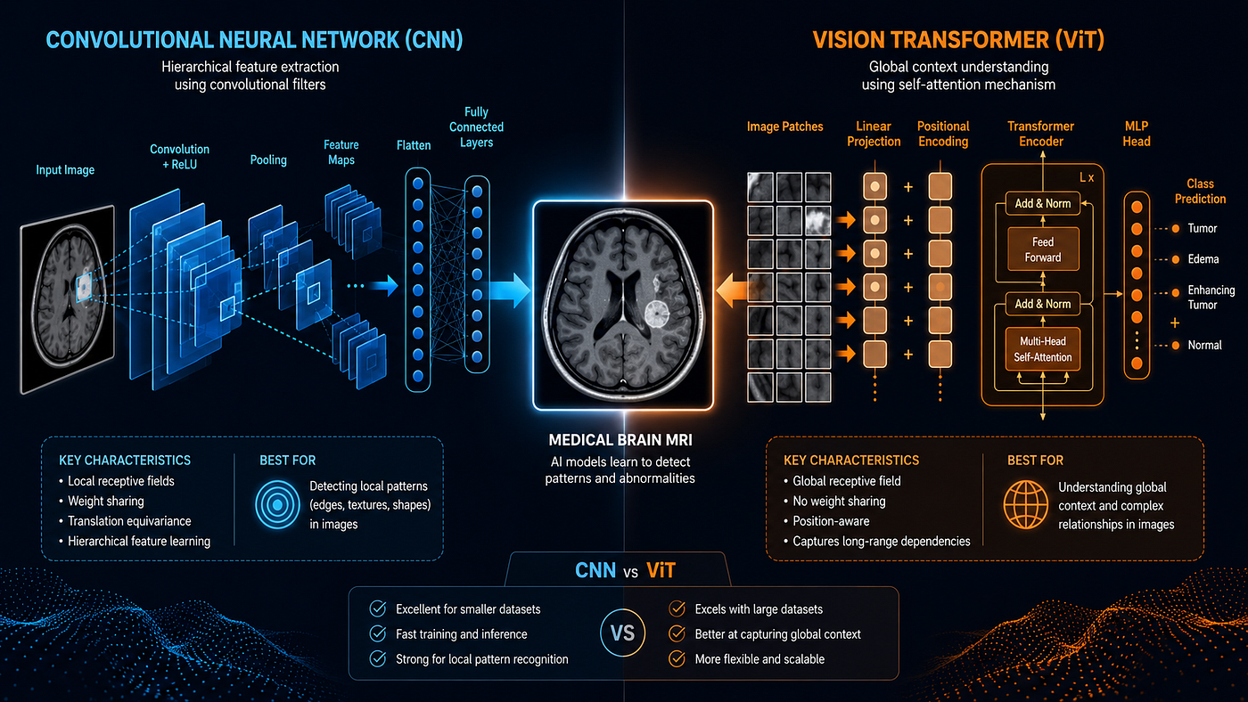
You have a medical imaging dataset. Maybe it is a collection of chest X-rays, MRI brain scans, or histopathology slides. You want to build a deep learning model that can classify, detect, or segment with high accuracy.
Now comes the question every ML practitioner and biomedical engineer eventually faces:
Which deep learning architecture should I actually use?
The answer is not as simple as picking the most popular model or the one with the highest benchmark score. Medical imaging has unique constraints limited labeled data, class imbalance, high stakes for errors, and strict interpretability requirements that make architecture selection a critical and nuanced decision.
This guide breaks it down clearly. By the end, you will know exactly how to evaluate and choose the right deep learning model for your medical imaging task.
Why Medical Imaging Is Different from Standard Computer Vision
Before diving into architectures, it is important to understand why medical imaging is a uniquely challenging domain.
1. Data scarcity. Unlike ImageNet with millions of labeled images, most medical datasets contain hundreds to a few thousand annotated samples. Overfitting is a constant risk.
2. High class imbalance. Diseased cases are often far outnumbered by healthy cases, requiring careful handling during training and evaluation.
3. Fine-grained features matter. The difference between a malignant and benign lesion can come down to subtle texture differences invisible at low resolution.
4. 3D data is common. CT scans and MRIs are volumetric standard 2D architectures do not naturally handle this.
5. Interpretability is non-negotiable. Clinical adoption requires that models explain their predictions, not just produce them.
These constraints directly influence which architectures are appropriate.
The Main Contenders: A Quick Overview
Here are the primary deep learning architectures used in medical imaging today:
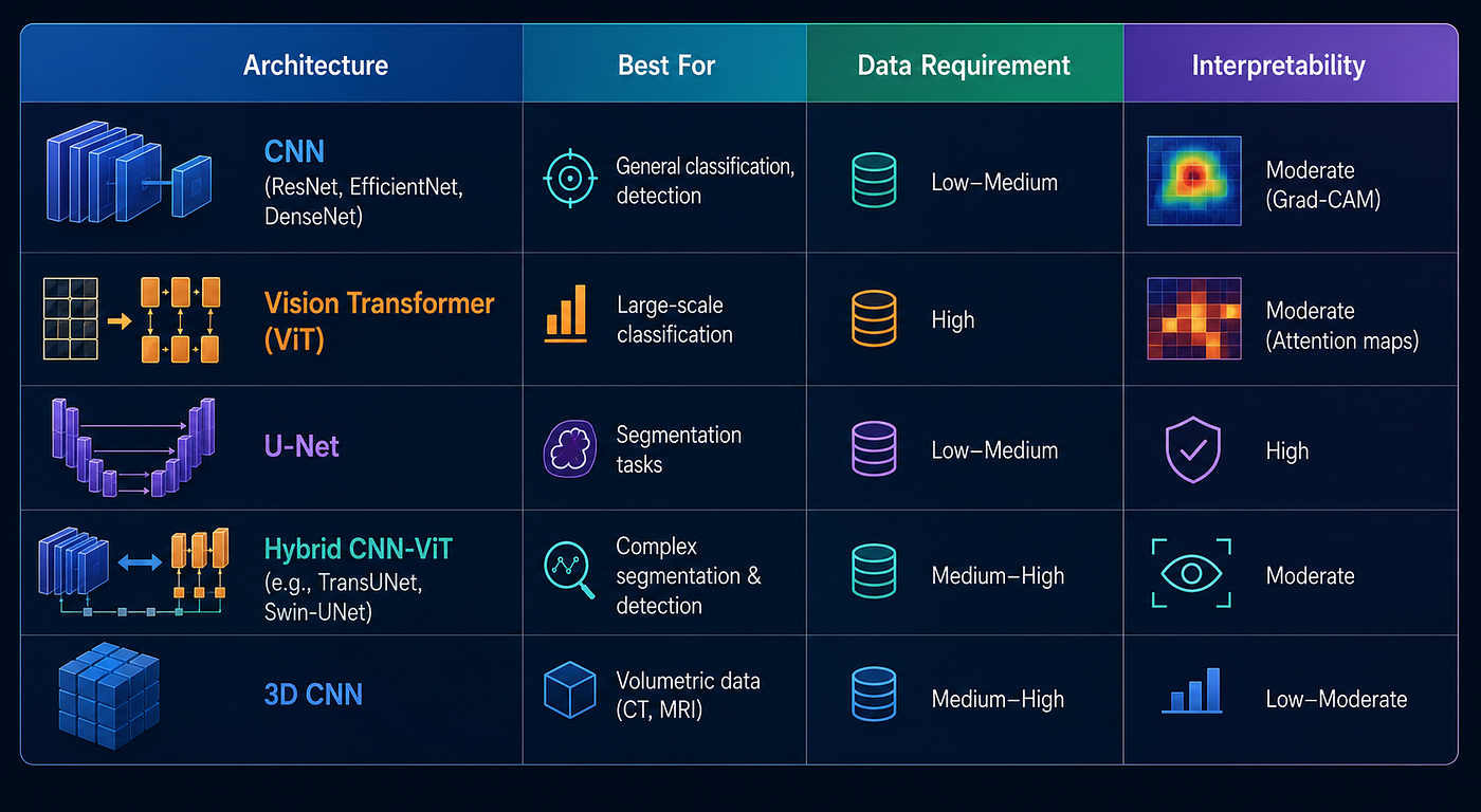
Option 1: CNNs The Reliable Workhorse
Convolutional Neural Networks remain the most widely deployed architecture in medical imaging, and for good reason.
Why CNNs Work Well in Medicine
CNNs are built to exploit spatial locality nearby pixels are more related than distant ones. This makes them naturally suited to detecting localized features in medical images: nodules in a chest CT, lesions in a retinal fundus image, or micro-calcifications in a mammogram.
They are also highly data-efficient compared to transformers. With transfer learning from ImageNet-pretrained weights (ResNet-50, EfficientNet-B4, DenseNet-121), a CNN can achieve strong performance with as few as a few hundred training images.
Best CNN Architectures for Medical Imaging
- ResNet-50 / ResNet-101 Reliable baseline for classification tasks. Residual connections prevent vanishing gradients in deep networks.
- Efficient Net (B0–B7) Excellent accuracy-to-parameter ratio. Strong choice when computational resources are limited.
- DenseNet-121 Used in the landmark CheXNet paper for chest X-ray pathology detection. Dense connections improve feature reuse, critical when labeled data is scarce.
When to Choose a CNN
- Your dataset has fewer than 10,000 labeled images
- You need a fast, deployable model with low computational cost
- Your task is 2D image classification or detection
- You need explain ability via Grad-CAM heatmaps
Option 2: Vision Transformers (ViT) The Powerful Newcomer
Vision Transformers apply the transformer architecture originally designed for NLP to image patches. Each image is divided into fixed-size patches, which are treated as tokens in a sequence.
Why ViT Is Compelling
ViTs capture long-range dependencies across an entire image in a way CNNs fundamentally cannot. For a CNN, understanding the relationship between a finding in the upper-left and one in the lower-right of an image requires many layers of computation. A ViT captures this relationship directly through its self-attention mechanism.
In medical imaging, this matters for tasks where global context is as important as local texture for example, understanding anatomical relationships in a full-body MRI or detecting diffuse disease patterns across an entire chest X-ray.
The Critical Limitation
ViTs are data-hungry. Standard ViT models were pre-trained on hundreds of millions of images (JFT-300M). Without sufficient data or strong pre-training, ViTs underperform CNNs significantly on small medical datasets.
This is being addressed by medical-domain pre-trained ViTs such as BioViL and MedViT, which are pre-trained specifically on biomedical image data.
When to Choose a ViT
- Your dataset is large (50,000+ images) or you have access to a medically pre-trained ViT
- Your task requires understanding global image context
- Computational resources are not a constraint
- You want attention map visualizations for explainability
Option 3: U-Net The Gold Standard for Segmentation
If your task is segmentation delineating the boundaries of an organ, tumor, or lesion U-Net is almost always the right starting point.
Why U-Net Dominates Medical Segmentation
Introduced in 2015 specifically for biomedical image segmentation, U-Net’s encoder-decoder architecture with skip connections allows it to capture both coarse spatial context (through downsampling) and fine-grained detail (recovered through upsampling and skip connections).
It was designed for small datasets and produces pixel-level predictions exactly what segmentation tasks require.
U-Net Variants Worth Knowing
- nnU-Net A self-configuring framework that automatically adapts preprocessing, architecture, and training for any segmentation dataset. Currently the state-of-the-art baseline for most segmentation benchmarks.
- Attention U-Net Adds attention gates to focus on relevant target regions, improving performance on small or subtle structures.
- 3D U-Net Extends U-Net to volumetric data for CT and MRI segmentation.
When to Choose U-Net
- Your task is semantic or instance segmentation
- You are working with limited labeled data
- You need pixel-level outputs (tumor masks, organ boundaries, lesion maps)
- You want a well-validated, clinically trusted architecture
Option 4: Hybrid CNN-ViT Models Best of Both Worlds
The latest frontier in medical imaging architecture combines the local feature extraction strength of CNNs with the global context modeling of transformers.
Key Hybrid Architectures
- TransUNet Combines a ResNet encoder with a transformer bottleneck and U-Net decoder. Strong performance on organ segmentation tasks.
- Swin-UNet A pure transformer-based U-Net that uses the Swin Transformer’s hierarchical, shifted-window attention. State-of-the-art on several medical segmentation benchmarks.
- MedT (Medical Transformer) Specifically designed for small medical datasets, using a gated axial-attention mechanism.
When to Choose a Hybrid Model
- You need both local texture sensitivity and global context understanding
- Your task is complex segmentation or multi-class detection
- You have a medium-to-large dataset and sufficient compute
- You want to push toward state-of-the-art performance
Option 5: 3D CNNs For Volumetric Medical Data
CT scans and MRIs are inherently three-dimensional. Processing them as a stack of 2D slices loses the spatial relationship between slices critical information for many diagnostic tasks.
3D CNNs extend standard convolutional operations into three dimensions, allowing the model to learn features across the full volume.
Trade-offs
3D CNNs are computationally expensive and require significantly more memory. They also need larger datasets to train effectively. Common approaches to manage this include:
- 2.5D methods Process three orthogonal 2D slices (axial, coronal, sagittal) through separate 2D CNNs and fuse the predictions.
- Patch-based training Train the 3D CNN on smaller volumetric patches rather than the full volume.
When to Choose a 3D CNN
- Your data is volumetric (CT, MRI, PET)
- Slice-to-slice context is critical for the diagnostic task
- You have sufficient compute and dataset size
Decision Framework: How to Choose
Use this step-by-step framework to select your architecture:
Step 1 — Define your task:
- Classification → CNN or ViT
- Segmentation → U-Net or hybrid
- Volumetric data → 3D CNN or 3D U-Net
Step 2 — Assess your dataset size:
- Fewer than 10,000 images → CNN with transfer learning or U-Net
- More than 50,000 images → ViT or hybrid CNN-ViT
Step 3 — Consider your compute constraints:
- Limited GPU → EfficientNet or standard U-Net
- High compute available → ViT, Swin-UNet, or 3D CNN
Step 4 — Evaluate interpretability needs:
- Clinical deployment required → Prefer CNN (Grad-CAM) or Attention U-Net
- Research setting → ViT attention maps acceptable
Step 5 — Start with a strong baseline: Always begin with a well-validated baseline (EfficientNet for classification, nnU-Net for segmentation) before exploring more complex architectures. Complexity rarely beats a well-tuned baseline on small medical datasets.
Quick Reference Summary
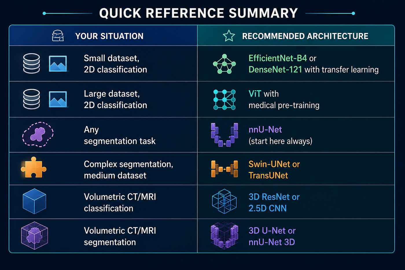
Final Thoughts
There is no universal “best” deep learning model for medical imaging. The right choice depends on your task, your data, your compute, and your deployment requirements.
What matters most is not picking the trendiest architecture it is understanding the strengths and limitations of each option and matching them to your problem. Start simple, validate rigorously, and add complexity only when your baseline demands it.
The best model is not the most sophisticated one. It is the one that is accurate, explainable, and trusted enough to actually help patients.
Recommended External Resources
- nnU-Net Framework (GitHub): https://github.com/MIC-DKFZ/nnUNet
- Papers With Code — Medical Image Segmentation Benchmarks: https://paperswithcode.com/task/medical-image-segmentation
- CheXNet Paper (Stanford ML Group): https://stanfordmlgroup.github.io/projects/chexnet/
- Hugging Face Medical Imaging Models: https://huggingface.co/models?pipeline_tag=image-classification&sort=downloads&search=medical
If this guide helped you make a better architecture decision, follow me on Medium for more practical deep learning and AI content.
메타데이터
- post_id
- 6d7cfd9074e2
- slug
- how-to-choose-the-best-deep-learning-model-for-medical-imaging-6d7cfd9074e2
- url
- https://medium.com/@rudradcruze/how-to-choose-the-best-deep-learning-model-for-medical-imaging-6d7cfd9074e2
- canonical_url
- https://medium.com/@rudradcruze/how-to-choose-the-best-deep-learning-model-for-medical-imaging-6d7cfd9074e2
- author_url
- https://medium.com/@rudradcruze
- status
- ok
- fetched_at
- 2026-07-21 01:40:58