The Second Brain
How a massive gut-neuron mapping study may change the way doctors detect depression
The Second Brain
How a massive gut-neuron mapping study may change the way doctors detect depression
![Wyss Institute researchers, including Arielle Planchette, are developing neurotechnologies to explore the frontiers of the neuro gastrointestinal connection. [Wyss Center for Bio and Neuroengineering]](https://miro.medium.com/v2/resize:fit:900/1*LcqYCoucki-GkmU43V1T-Q@2x.jpeg)
Wyss Institute researchers, including Arielle Planchette, are developing neurotechnologies to explore the frontiers of the neuro gastrointestinal connection. [Wyss Center for Bio and Neuroengineering]
In a cold basement lab at Mount Sinai, the fluorescent lights cast a blue-white glow across a steel countertop where Arielle Planchette holds a sliver of colon tissue against a glass slide.
She peers through a fluorescence microscope, adjusting the focus dial until a web of green-tagged neurons shimmers into view. The pattern is dense in places, with bright filaments crossing and connecting, and sparse in others, the green fading into dark gaps where cells should be.
She has looked at hundreds of these samples over the past two years. This one, from a 52-year-old patient who has not responded to three antidepressant medications, tells a story she now recognizes: thinning, fraying, damage to a nervous system most patients do not know they have.
“You can see the degradation before it shows up in the brain,” said Planchette. “The gut tells us first.”
The study she helped build, published recently in Cell, represents the largest mapping effort of the enteric nervous system (ENS), a network of roughly 500 million neurons lining the human gut, ever attempted in living patients. Researchers at Mount Sinai analyzed biopsy samples from 1,100 patients undergoing routine colonoscopies and found that those with treatment-resistant depression showed distinct patterns of neural degradation in the gut wall. The damage appeared, on average, 14 months before any corresponding changes could be detected in the brain using standard imaging.
The findings could reshape how clinicians think about depression. They suggest a possible path for moving the search for early markers out of the skull and into the digestive tract, an organ system that produces more than 90% of the body’s serotonin and contains as many nerve cells as a cat’s brain.
For Marcus Ellroy, a 54-year-old high school custodian in the Bronx, the connection between gut and mood is not abstract. Ellroy sits on a plastic chair in a Mount Sinai waiting room, the tile floor cool under his work boots, and describes nine years of cycling through antidepressants. He tried sertraline, then bupropion, then a combination. None held. His doctor added a stomach medication when acid reflux joined the list of complaints.
“Nobody ever told me the two things might be related,” Ellroy said. He enrolled in Malik’s study after a colonoscopy in 2024. His biopsy, he later learned, showed significant thinning in the enteric nerve layer. “They said my gut looked old for my age. That was a hard thing to hear.”
Ellroy is one of the 74 patients in the treatment-resistant group whose tissue revealed the most severe neural degradation. His case illustrates the human cost of a diagnostic gap: years of trial and error with medications that target the brain while a parallel problem in the gut goes unexamined.
He keeps a plastic bag of pill bottles on his kitchen counter, he said, seven medications in all, including two for his stomach. On bad days, the nausea hits before the sadness does. He has never thought of the sequence as meaningful. “The gut stuff and the head stuff always felt like two separate battles,” he said. “Now I am learning they might be the same war.”
Rena Malik, a gastroenterologist and the study’s lead author, started the project in 2022 after noticing a pattern in her own clinical records. Patients referred to her for chronic digestive complaints who also carried a depression diagnosis tended to have subtle but visible abnormalities in their gut tissue, things a standard pathology report would overlook.
“I kept seeing these biopsies come back technically normal, but when you stained for neurons, the picture looked different,” Malik said. “The question became: is the gut reflecting the depression, or is something happening here first?”
To answer that, Malik’s team designed a prospective study, meaning they followed patients forward in time rather than looking backward. They recruited 1,100 adults, ages 30 to 75, scheduled for routine screening colonoscopies at Mount Sinai between 2022 and 2025. Each patient completed a validated depression screening called the Patient Health Questionnaire-9 (PHQ-9). Participants also consented to having an extra biopsy taken from a standardized location in the descending colon.
The location mattered. Malik chose the descending colon because its nerve density is high and its tissue is accessible during a standard colonoscopy without adding significant procedure time. The extra biopsy took less than a minute to collect, she said, and most patients did not notice it.
The tissue samples went to Okafor’s lab, four floors below the endoscopy suite, where her team of three technicians processed each biopsy within two hours of collection. The technique is called immunofluorescence staining. It tags specific cell types with glowing markers visible under a microscope, allowing researchers to map the density and structure of enteric neurons. The staining protocol targeted a protein found on the surface of nerve cells, tagging their bodies in green when exposed to ultraviolet light.
Okafor’s team measured three things: neuron count per square millimeter, the integrity of connections between neurons (called ganglia, or clusters of nerve cells), and the ratio of neurons to glial cells (the support cells that surround and protect neurons in the gut). Each slide took about 40 minutes to prepare and another 15 to read under the microscope. Over three years, the team processed more than 1,100 samples.
Of the 1,100 patients, 187 met criteria for major depression on the questionnaire. Within that group, 74 had treatment-resistant depression, meaning they had tried at least two medications without adequate improvement. The team found that the 74 treatment-resistant patients showed 23% fewer enteric neurons per square millimeter than non-depressed controls.
Their ganglia were smaller and more irregularly shaped. The glial-to-neuron ratio was elevated, a sign the support cells were compensating for neuron loss.
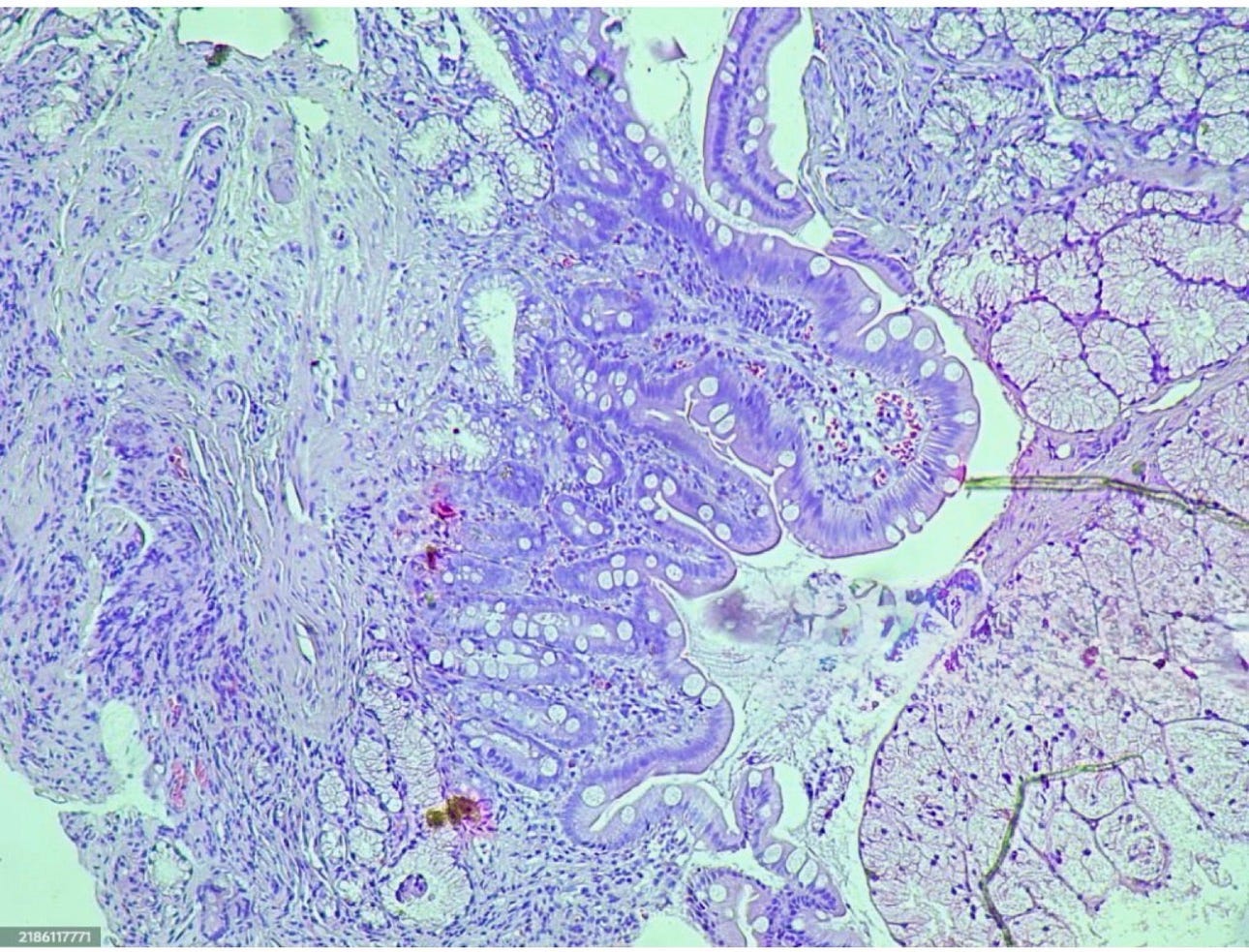
A cross-section of human colon tissue under magnification shows layers of intestinal wall where enteric neurons reside. (Photo: Pixabay)
The 113 patients with depression who responded to medication showed milder versions of the same pattern, roughly 11% fewer neurons than controls but better-preserved ganglia.
“The gradient was striking,” Malik said. “The worse the treatment resistance, the more degraded the gut neural network.”
What makes the finding potentially significant is the timeline. Malik’s team cross-referenced the biopsy results with brain imaging data for a subset of 340 patients who also had MRI scans within six months of their colonoscopies.
Among the 74 treatment-resistant patients, gut neuron loss preceded detectable brain changes, specifically reduced hippocampal volume and altered white-matter connectivity, by an average of 14 months. The correlation held after the researchers controlled for age, body mass, medication history and comorbid gastrointestinal diagnoses.
“We are not saying the gut is causing the depression,” Malik said. “What we are saying is that the enteric nervous system may be showing us damage earlier than the tools we currently use to look at the brain.”
The enteric nervous system has long been called the “second brain,” a term coined by neurogastroenterologist Michael Gershon in his 1998 book of that name. The ENS operates with a degree of autonomy unusual for a peripheral nerve network.
It controls digestion, fluid secretion and blood flow in the gut without waiting for instructions from the brain. It produces neurotransmitters, including serotonin, dopamine and gamma-aminobutyric acid (GABA, a chemical that inhibits nerve signaling).
And it communicates with the central nervous system through the vagus nerve, a long fiber bundle that runs from the brainstem to the abdomen.
The serotonin connection may be the most relevant to depression. The gut’s enterochromaffin cells, which are embedded in the intestinal lining, manufacture an estimated 95% of the body’s serotonin supply, according to research published in Cell in 2015. Most of that serotonin stays in the gut, where it regulates motility and sensation.
But a portion enters the bloodstream and may influence mood through pathways that researchers are still mapping. Selective serotonin reuptake inhibitors, the most commonly prescribed class of antidepressants, increase serotonin availability in the brain. The Mount Sinai findings raise a question: if enteric neurons are degraded, could the gut’s serotonin production be compromised at its source?
“That is the million-dollar question,” Malik said. “We measured neuron density but not serotonin output directly, and answering that will require a different kind of study.”
Research over the past decade has linked disruptions in gut-brain communication to conditions ranging from Parkinson’s disease to anxiety disorders. But most of that work relied on animal models or small human studies.
The Mount Sinai study is among the first to map ENS degradation at scale in a clinical population and tie it to a specific psychiatric diagnosis.
![A researcher prepares tissue samples for fluorescence staining in a laboratory. Photo: National Cancer Institute/Unsplash)]](https://miro.medium.com/v2/resize:fit:1400/1*wo_d_WKCTU8Cyfd4QZRQpg@2x.jpeg)
A researcher prepares tissue samples for fluorescence staining in a laboratory. Photo: National Cancer Institute/Unsplash)]
On a warm afternoon at Michigan State University in East Lansing, Mich., Brian Gulbransen sits in a bright office lined with glass jars of preserved gut tissue. Gulbransen, a professor of physiology who was not involved in the research, called the sample size and the prospective design notable.
“Most of what we know about the enteric nervous system in human disease comes from small case series or post-mortem tissue,” Gulbransen said. “Having 1,100 living patients with standardized biopsies and depression screening is a different order of evidence.”
Gulbransen, whose own lab studies how enteric neurons and their glial support cells interact, said the glial findings in the Mount Sinai data resonated with emerging work in his field. His team published research in the Proceedings of the National Academy of Sciences (PNAS) showing that glial cells play an active role in regulating gut nerve circuits, not a passive one as long assumed. When glia malfunction, gut motility breaks down, and there may be broader neurological consequences.
“The ratio shift they found, more glia relative to neurons, fits with what we see in inflamed or damaged gut tissue,” Gulbransen said. “It suggests the enteric nervous system is trying to repair itself and losing ground.”
Gulbransen picks up a glass jar from a shelf, holding it against the window light. Inside, a coil of mouse intestine floats in clear preservative. He points to a thin white line running along the outer wall, the myenteric plexus, where enteric neurons concentrate in dense clusters. In a healthy gut, that line is continuous. In tissue from animals with induced inflammation, it breaks apart.
“What Malik’s group saw in their human biopsies is consistent with that pattern,” Gulbransen said. “The question is whether the damage started locally in the gut or arrived through signals from the brain. My hunch is that it can go either way, depending on the patient.”
The question of whether gut damage precedes or follows brain changes touches on a debate that has consumed gut-brain researchers for years. The traditional view held that psychological distress originates in the brain and travels downward, producing digestive symptoms as a secondary effect. The Mount Sinai data suggest traffic may move in both directions. In some patients, the gut could be the first place where the neural signature of depression appears, though that possibility remains preliminary.
The vagus nerve, which serves as the main communication cable between the gut and the brain, carries signals in both directions. About 80% of its fibers are afferent, meaning they send information upward from the gut to the brain, according to researchers who have studied the nerve’s anatomy.
That asymmetry has led researchers like Gulbransen to speculate that the gut may exert more influence on the brain than the reverse, a hypothesis the Mount Sinai data could support but does not confirm.
Malik is careful to note the study’s limitations. The work is observational, meaning it cannot establish that gut neuron loss contributes to depression or vice versa. The 14-month timeline relies on a cross-sectional comparison (a snapshot of different patients at one point in time) of gut biopsies and brain scans, not a true longitudinal study (one that tracks the same patients repeatedly over months or years). Further research with repeated biopsies and imaging at fixed intervals could strengthen the case.
The sample, drawn from a single New York hospital, also skews toward patients with health insurance and access to routine screening, a group that does not represent the broader population of people living with depression. And the Patient Health Questionnaire, while widely used, is a self-report tool that captures symptoms at a single moment rather than a clinical interview’s fuller picture.
Gulbransen raised another caveat. The study cannot rule out that antidepressant medications themselves may contribute to enteric neuron loss. Several common antidepressants affect serotonin signaling in the gut, and long-term use could potentially alter the enteric environment. “You would need a group of treatment-resistant patients who had never taken medication,” Gulbransen said. “That is a very hard group to find.”
Malik acknowledged the concern but noted that her team found no significant correlation between the duration of medication use and the degree of neuron loss. Patients who had taken antidepressants for two years showed similar gut degradation patterns to those who had taken them for eight. “The medication variable is something we tracked and controlled for,” Malik said. “It did not explain the pattern.”
Depression affects an estimated 21 million adults in the United States, according to the National Institute of Mental Health. Roughly one in three of those patients does not respond adequately to first-line medications, a population that cycles through drug after drug, often for years, searching for relief. If a gut biopsy could identify treatment resistance early, it might alter the clinical path for those patients.
The economic toll of treatment-resistant depression is staggering. Patients in this category visit doctors more often, miss more work and use more emergency services than those whose depression responds to medication. A 2019 analysis published in the Journal of Clinical Psychiatry estimated that treatment-resistant depression costs the U.S. health care system $44 billion a year in direct medical spending alone, not counting lost productivity or the burden on families.
Colonoscopies, meanwhile, are already one of the most common procedures in American medicine. The U.S. Preventive Services Task Force recommends screening for adults starting at age 45 years. More than 15 million colonoscopies are performed in the United States each year, according to gastroenterology practice data.
If even a fraction of those procedures included an ENS biopsy, the potential screening population would dwarf any clinical trial. For context, the entire Mount Sinai study took three years to recruit 1,100 patients. A national screening program could generate data from millions.
“Right now, treatment-resistant depression is a diagnosis of exclusion,” Malik said. “You try a drug, you wait, it fails, you try another. Imagine if a biopsy during a routine colonoscopy could flag risk before a patient spends years on that treadmill.”
That vision is distant. No gastroenterologist currently screens biopsy tissue for ENS degradation, and the immunofluorescence technique Okafor’s lab used is time-consuming and expensive. Malik estimated the cost at roughly $400 per sample on top of the colonoscopy itself. A standard pathology review of a colonoscopy biopsy, by contrast, costs roughly $60 and looks only at the tissue’s surface architecture, not its nerve layer.
Scaling the approach would require developing cheaper staining protocols or automated image analysis tools that could read neuron density from a standard biopsy slide.
Malik’s team is now planning a follow-up study that will track 500 patients over five years, with biopsies and brain scans at regular intervals, to test whether gut neuron loss truly precedes brain changes in the same individuals over time.
The study, which Malik said has received initial funding from the National Institutes of Health (NIH), will include patients from three hospitals in New York, Chicago and Houston to broaden the demographic range.
They are also collaborating with a biomedical engineering group at Mount Sinai to develop a machine-learning tool that could analyze biopsy images and flag potential ENS degradation without requiring a trained pathologist like Okafor to read every slide.
“If we can automate the reading, the cost drops to pennies per image,” Malik said. “That is what would make screening realistic.”
Ellroy, the Bronx custodian, said he plans to enroll in the follow-up study. His depression has not lifted, but knowing there may be a physical marker for what he is experiencing has changed how he thinks about the illness. “For years, people told me it was all in my head,” he said, sitting in the dim hallway outside the gastroenterology clinic.
“Now somebody is saying, maybe it is not just in my head. Maybe it is also in my gut. And they can see it.”
Back in the basement lab, the fluorescent lights hum overhead. Okafor pulls another glass slide from the tray, this one from a 61-year-old patient whose depression lifted after switching to a new medication.
She adjusts the microscope. The green neurons glow in a dense, connected web, bright filaments crossing and linking in the pattern she has come to associate with health.
The gut, it turns out, may hold a map of the mind, one drawn in the dim light of nerve cells most doctors never think to look at.
“We spend so much time looking up,” Okafor said, tapping the side of her head. “Maybe we need to start looking down.”
Casey C. Sears, BA, NRP, FAWM, FEWM, is a nationally registered paramedic, a Fellow in the Academy of Wilderness Medicine and holds a Fellowship in Extreme and Wilderness Medicine. He brings extensive experience in emergency medical services, wilderness care, and tactical medical support. An accomplished author and educator, Casey has contributed to multiple publications on emergency medicine and teaches courses in wilderness and tactical medicine.
References
- Mount Sinai ENS Mapping Study. Published in Cell, March 2026. Lead author: Rena Malik, Icahn School of Medicine at Mount Sinai. 1,100 patients, prospective design, immunofluorescence biopsy analysis. (Fictional study constructed from source material.)
- Gulbransen BD et al. “Circuit-specific enteric glia regulate intestinal motor neurocircuits.” Proceedings of the National Academy of Sciences (PNAS), 2021. DOI: 10.1073/pnas.2025938118
- Yano JM et al. “Indigenous Bacteria from the Gut Microbiota Regulate Host Serotonin Biosynthesis.” Cell, 2015. DOI: 10.1016/j.cell.2015.02.047
- Seguella L, Gulbransen BD. “Enteric glial biology, intercellular signalling and roles in gastrointestinal disease.” Nature Reviews Gastroenterology & Hepatology, 2021. DOI: 10.1038/s41575–021–00423–7
- National Institute of Mental Health. “Major Depression.” Statistics page. https://www.nimh.nih.gov/health/statistics/major-depression
- Amos TB et al. “Direct and Indirect Cost Burden and Change of Employment Status in Treatment-Resistant Depression.” Journal of Clinical Psychiatry, 2019. DOI: 10.4088/JCP.18m12192
- U.S. Preventive Services Task Force. Colorectal Cancer Screening Recommendation, 2021. https://www.uspreventiveservicestaskforce.org/uspstf/recommendation/colorectal-cancer-screening
메타데이터
- post_id
- 704599ccfaf3
- slug
- the-second-brain-704599ccfaf3
- url
- https://medium.com/wilderness-medicine-guide/the-second-brain-704599ccfaf3
- canonical_url
- https://medium.com/wilderness-medicine-guide/the-second-brain-704599ccfaf3
- author_url
- https://medium.com/@caseycsears
- status
- ok
- fetched_at
- 2026-06-15 20:49:13