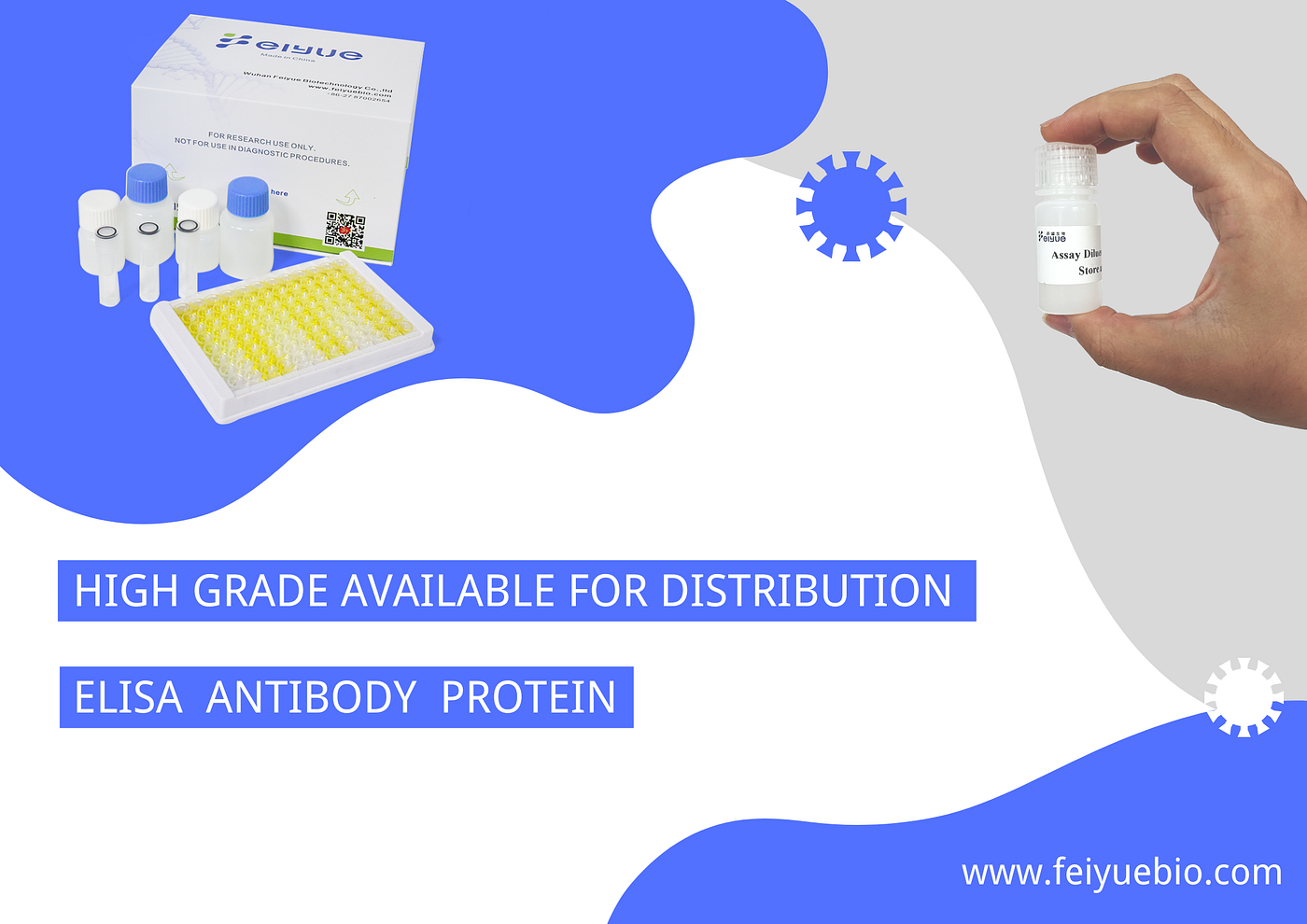
Human Tau Protein (MAPT) ELISA kit
Catalog No:FY-EH6846
Human Tau Protein (MAPT) ELISA kit
Catalog No:FY-EH6846
Size: 96T
Intended Use
The kit is intended for use in quantitative determination of Human MAPT concentrations in serum, plasma, tissue homogenates, cell lysates, cell culture supernates and other biological fluids.
Specification
Sensitivity: 18.75 pg/mL.
Detection Range: 31.2–2000 pg/mL.
No significant cross-reactivity or interference between Human MAPT and analogues was observed.
Repeatability: Coefficient of variation is < 10%.
Principle of the Procedure
This kit uses the Sandwich-ELISA principle. The microtiter plate strips has been pre-coated with an affinity purified antibody to Human MAPT. Standards or samples containing Human MAPT are added to the plate and reacted with capture antibody. A second anti-Human MAPT antibody labeled biotin is then added and binds to Human MAPT captured on the plate. After that, Streptavidin-Horseradish Peroxidase(SA-HRP) is added to form a sandwich complex of solid phase antibody-Human MAPT-biotin labeled antibody-SA-HRP. And then, TMB substrate solution is added to all wells and incubated. An enzyme-catalyzed reaction generates a blue color in the solution, thereafter, stop solution is added to stop the substrate reaction and the color turns yellow. The yellow solution is read at a wavelength of 450nm. The concentration of Human MAPT in the samples is then calculated from the OD value by establishing a standard curve.
Limitations of the Procedure
-
This kit is for laboratory scientific research only, we will not be responsible for any consequences if this kit is used for clinical diagnosis or any other procedures.
-
Due to the uncertainty of its validity, this kit may not be suitable for testing some special experimental samples, such as gene knockout experiments.
-
This kit should be used before its expiration date, and please strictly follow the instructions for storage.
-
Different manufacturers’ kits or testing the same analyte by other methods may produce inconsistent results because we do not compare our products with those of other manufacturers.
-
Since the antibodies used in the kit are usually prepared from recombinant proteins as immunogen, and recombinant proteins can be limited by different fragmentation, expression and purification systems, it is not recommended to use this kit to detect recombinant proteins
-
In order to get the best experimental results, please use only the reagents provided by the manufacturer, and do not mix reagents from different batches.
-
Due to the existing conditions and the limitations of science and technology, we cannot fully identify and analyze the raw materials provided by the supplier comprehensively. Therefore, the kit may have some quality and technical risks.
-
The possibility of interference cannot be excluded before all factors are tested in the ELISA immunoassay.
-
In order to obtain reproducible results, each step in the experiment should be controlled and variations in sample collection, handling and storage may also lead to differences in sample measurements.
10.Although each kit passes rigorous quality testing, differences in measured values between batches of kits can still be caused by factors such as shipping conditions and different laboratory equipment.
Storage & Stability
-
After receiving the kit, an unopened kit can be stored at 2–8℃ for 1 month. If the kit is not used within 1 month, store each component separately at the temperature indicated in the table above once the kit is received.
-
When the kit is used, the remaining reagents should be stored according to the table above storage condition.Unused plate strips should be immediately returned to the foil bags containing the desiccant, resealed and stored at -20℃.
-
All reagent bottle caps must be tightened to prevent evaporation and microbial pollution, the volume of imeasuring equipment instead of directly pouring into the vials.
-
The expiration date of the product is determined by the label on the box, and all components are guaranteed to be stable during the shelf life.
Materials & Equipment Required But Not Provided
Microplate reader with 450nm wavelength filter
Incubator capable of maintaining 37℃
Single or multi-channel pipettes with high precision
disposable pipette tips
EP tubes
Container for Wash Solution
Squirt bottle, manifold dispenser, or automated microplate washer
Deionized or distilled water
Absorbent paper for blotting the microplate
Note for Procedure
-
Please wear lab coats, eye protection and latex gloves for protection. Please perform the experiment following the national security protocols of biological laboratories, especially when detecting blood samples or other bodily fluids.
-
A freshly opened ELISA Plate may appear to have a water-like substance, which is normal and ill not have any impact on the experimental results.
-
Do not reuse the reconstituted standard, Detection Reagent A working solution, Detection Reagent B working solution.
-
The microplate reader should have a 450(±10 nm) filter installed and a detector that can detect the wavelength. The optical density should be within 0–3.5.
-
The instruction manual is also for the kit of 48T, but all reagents of 48T kit are reduced by half.
-
When mixing or reconstituting protein solutions, always avoid foaming.
-
To avoid cross-contamination, change pipette tips between additions of each standard level, between sample additions, and between reagent additions. Also, use separate reservoirs for each reagent.
-
To ensure accurate results, proper adhesion of plate sealers during incubation steps is necessary.
-
When using an automated plate washer, adding a 30 second soak period following the addition of Wash Buffer, between wash steps may improve assay precision.
-
Substrate Solution should remain colorless until added to the plate. Keep Substrate Solution protected from light. Substrate Solution should change from colorless to gradations of blue.
-
Stop Solution should be added to the plate in the same order as the Substrate Solution. The color developed in the wells will turn from blue to yellow upon addition of the Stop Solution. Wells that are green in color indicate that the Stop Solution has not mixed thoroughly with the Substrate Solution.
Sample collection
Serum: Allow blood samples to clot for 2 hours at room temperature or overnight at 2–8℃ before centrifugation for 15 min at 2000×g at 2–8℃. Supernatants should be taken for assay testing immediately or stored at -20℃ or -80℃ for later use.
Plasma: Collect plasma using EDTA or heparin as an anticoagulant. Centrifuge samples for 15 min at 2000×g at 2–8℃ within 30 min of collection. Supernatants should be taken for assay testing immediately or stored at -20℃ or -80℃ for later use.
Tissue homogenates: Tissues should be rinsed in cool PBS to remove excess blood thoroughly, weighed, minced into small pieces. then Tissue pieces omogenized in cool PBS (tissue weight (g): PBS (mL) volume=1:9) with a glass homogenizer on ice. The resulting suspension is sonicated with an ultrasonic cell disrupter till the solution is clarified. The homogenates are then centrifuged for 5 min at 10000×g. Supernatants should be taken for assay testing immediately or stored at -20℃ or -80℃ for later use.
Cell lysates: For adherent cells, gently wash the cells with moderate amount of cool PBS and detach the cells with trypsin. Collect cells by centrifugation for 5 min at 1500×g(suspension cells can be collected by centrifugation directly). Discard the supernate and wash cells 3 times with cool PBS. Resuspend cells in cool PBS with concentration of 5×10⁶ cells/mL. Repeat the freeze-thaw process several times until the cells are fully lysed. Centrifuge for 15min at 2000×g at 2–8℃. Supernatants should be taken for assay testing immediately or stored at -20℃ or -80℃ for later use.
Cell culture supernatant and other biological fluids: Centrifuge samples for 15 min at 1500×g at 2–8℃. Supernatants should be taken for assay testing immediately or stored at -20℃ or -80℃ for later use.
Note for sample
-
Samples should be used within 6 days when stored at 2–8℃, otherwise samples must be stored at -20℃ (≤1month) or -80℃ (≤2months). Avoid repeated freeze-thaw cycles.
-
Please predict the concentration before assaying. If the sample concentration is not within the range of the standard curve, users must determine the optimal sample dilutions for their particular experiments.
-
If the samples are not indicated in the manual, a preliminary experiment to determine the validity of the kit is necessary.
-
If a lysis buffer is used to prepare tissue homogenates or cell culture supernatant, there is a possibility to cause a deviation due to the introduced chemical substance.
-
Some recombinant protein may not be detected due to a mismatching with the coated antibody or detection antibody.
-
Please do not use hemolyzed samples for ELISA as it will affect the test results.
-
Fresh samples without long time storage is recommended for the test. Otherwise, protein degradation and denaturalization may occur in those samples and finally lead to wrong result.
Reagent Preparation
-
Allow all reagents to return to room temperature (18–25℃) before use.
-
Wash Buffer: Dilute 20 mL of Concentrated Wash Buffer with 580 mL of deionized or distilled water to prepare 600 mL of Wash Buffer.
-
Standard Working Solution: First, centrifuge the standard at 1000×g for 1 min, add 1mL of standard sample dilution, let it stand for 10min and then mix gently, that is 8000pg/mL of standard working solution.
Second, take 8 EP tubes, add 500μL of Standard & Sample Diluent to each tube. Pipette 250μL of the 8000pg/mL working solution and 250μL of Standard & Sample Diluent to the first tube and mix up to produce a 2000pg/mL working solution. Pipette 500μL of the solution from the former tube into the latter one according to the picture shown below. Mix each tube thoroughly before the next transfer. Set up 7 points of diluted standard such as 2000pg/mL, 1000pg/mL, 500pg/mL, 250pg/mL, 125pg/mL, 62.5pg/mL, 31.2pg/mL, and the last EP tubes with Standard & Sample Diluent is the blank as 0pg/mL.

-
Detection Reagent A working solution: Calculate the required amount before the experiment (100 μL/well). In preparation, slightly more than calculated should be prepared. Centrifuge the stock tube before use, dilute the 100× Concentrated Detection Reagent A to 1×working solution with Assay Diluent A.
-
Detection Reagent B working solution: Calculate the required amount before the experiment (100 μL/well). In preparation, slightly more than calculated should be prepared. Centrifuge the stock tube before use, dilute the 100× Concentrated Detection Reagent B to 1× working solution with Assay Diluent B.
Assay Protocol
- Add the Standard working solution to the first two columns: Each concentration of the solution is added in duplicate, to one well each, side by side (100 μL for each well). Add the samples to the other wells (100 μL for each well). Cover with the plate sealer provided in the kit. Incubate for 60 min at 37℃.
2. Discard the liquid from each well, don’t wash. Immediately add 100μL of Detection Reagent A working solution to each well. Cover with the plate sealer. Incubate for 60 min at 37℃.
-
Discard the liquid from each well, add 350μL of wash buffer to each well. Soak for 30sec and decant the solution from each well and pat it dry against clean absorbent paper. Repeat this wash step 3 times in total.
-
Add 100μL of Detection Reagent B working solution to each well. Cover with the plate sealer. Incubate for 30 min at 37℃.
-
Turn on the enzyme marker for preheating.
-
Discard the liquid from each well, repeat the washing process of step 3 for 5 times.
-
Add 90μL of TMB Reagent to each well. Cover with a new plate sealer. Incubate for 10–20min at 37℃. Protect the plate from light.
-
Add 50 μL of Stop Solution to each well.
-
The absorbance OD value of each well is measured at 450nm right away.
Calculation of Results
Average the duplicate readings for each standard and samples, then subtract the average zero standard optical density. Create a standard curve with standard concentration on the x-axis and OD values on the y-axis. Draw a best fit curve through the points and it can be determined by regression analysis. If samples have been diluted, the concentration read from the standard curve must be multiplied by the dilution factor.

Sensitivity
The minimum detectable dose of Human MAPT is typically less than 18.75pg/mL.
The sensitivity of this assay, or Limit of Detection (LOD) was defined as the lowest protein concentration that could be differentiated from zero. It was determined by adding three standard deviations to the mean OD value of twenty zero standard replicates and calculating the corresponding concentration.
Specificity
This assay has high sensitivity and excellent specificity for detection of Human MAPT.
No significant cross-reactivity or interference between Human MAPT and analogues was observed.
Limited by current skills and knowledge, it is impossible for us to complete the cross- reactivity detection between Human MAPT and all the analogues, therefore, cross reaction may still exist.
Summary for Procedure
-
Prepare all the reagents
-
Add 100μL Standard or Sample. Incubate 60mintues at 37℃
-
Discard the liquid. Add 100μL Detection Reagent A
Incubate 60mintues at 37℃
-
Aspirate and wash 3 times
-
Add 100μL Detection Reagent B. Incubate 30 minutes at 37℃
-
Aspirate and wash 5 times
-
Add 90μL TMB Reagent. Incubate 15 minutes at 37℃
-
Add 50μL Stop Solution
-
Read at 450nm immediately and calculate of results
메타데이터
- post_id
- 8db2e1a2b925
- slug
- human-tau-protein-mapt-elisa-kit-8db2e1a2b925
- url
- https://medium.com/@czy15701/human-tau-protein-mapt-elisa-kit-8db2e1a2b925
- canonical_url
- https://medium.com/@czy15701/human-tau-protein-mapt-elisa-kit-8db2e1a2b925
- author_url
- https://medium.com/@czy15701
- status
- ok
- fetched_at
- 2026-06-18 00:10:23