After the Heart Stops
A silent alarm in the open-heart theater
After the Heart Stops
A silent alarm in the open-heart theater
Operating rooms carry a peculiar kind of calm. It is the stillness you find at the eye of a storm, laced with disinfectant, punctuated by monitor beeps, and threaded with a team’s wordless choreography. On this day the mitral valve replacement unfolds like a well-rehearsed symphony. The surgeon’s hands move with unhurried grace. The final stitch sets, the paddles deliver a jolt, and the heart returns to order. Curves on the screen gather themselves, regular and reassuring. We breathe out. Another life, pulled back from the ledge.
Then vision narrows to a small detail at the bed’s edge. The Foley catheter hangs, a clear conduit to a limp collection bag. It should hold a warm pool of gold. Instead, it is slack and empty.
In the surgeons’ moment of victory, the ICU’s battle begins. The dry urine bag is a quiet siren, a hint that while the heart has been restored, another organ is staging a strike. This is cardiac surgery–associated acute kidney injury, CSA-AKI for short, a silent adversary that lurks in the shadow of every triumphant “open heart.”
Opening the heart, opening a box
The history of cardiac surgery reads like a string of impossible feats made possible. Yet for all the courage it took to tame the heart, the kidney paid a price. That price is not a footnote. Even in the modern era, thirty to forty percent of patients experience some degree of AKI after cardiac surgery, and a smaller but consequential fraction slide into severe injury, dialysis, and death. The story is not about numbers alone. It is about genius and stubbornness, accidental harms and sober reckonings, the halting progress of medicine as it stumbles from “who knew” to “now we know.” It begins with a stopped heart and a machine that sounded like madness.
Dr. Gibbon and his audacious machine
John H. Gibbon Jr., born into a dynasty of Philadelphia physicians, nearly traded medical school for poetry. His father talked him into staying, and the world’s hearts are different because of it.
The idea that gripped him did not arrive as inspiration so much as desperation. In 1931 he watched a woman with a massive pulmonary embolism teeter at the edge of death for seventeen hours. He imagined taking her dark, oxygen-starved venous blood out of the body, filling it with oxygen, washing off carbon dioxide, and sending it back red and bright. If only a machine could do this, he thought, she might live.
Colleagues dismissed the proposal as a glamorous way to waste a career. Gibbon found an ally in his wife, Mary “Maly” Hopkinson Gibbon, a trained researcher who became his closest collaborator. Their lab looked more like a salvage yard than a temple of science. Early heart-lung rigs were rubber, glass, scrap metal, and homemade valves. Money was scarce; nights were spent trapping stray cats to keep the animal studies going. Out of this improvised workshop, their contraption kept a cat alive after the heart had been motionless for nearly thirty minutes, a victory Gibbon later said surpassed any other joy of his career.
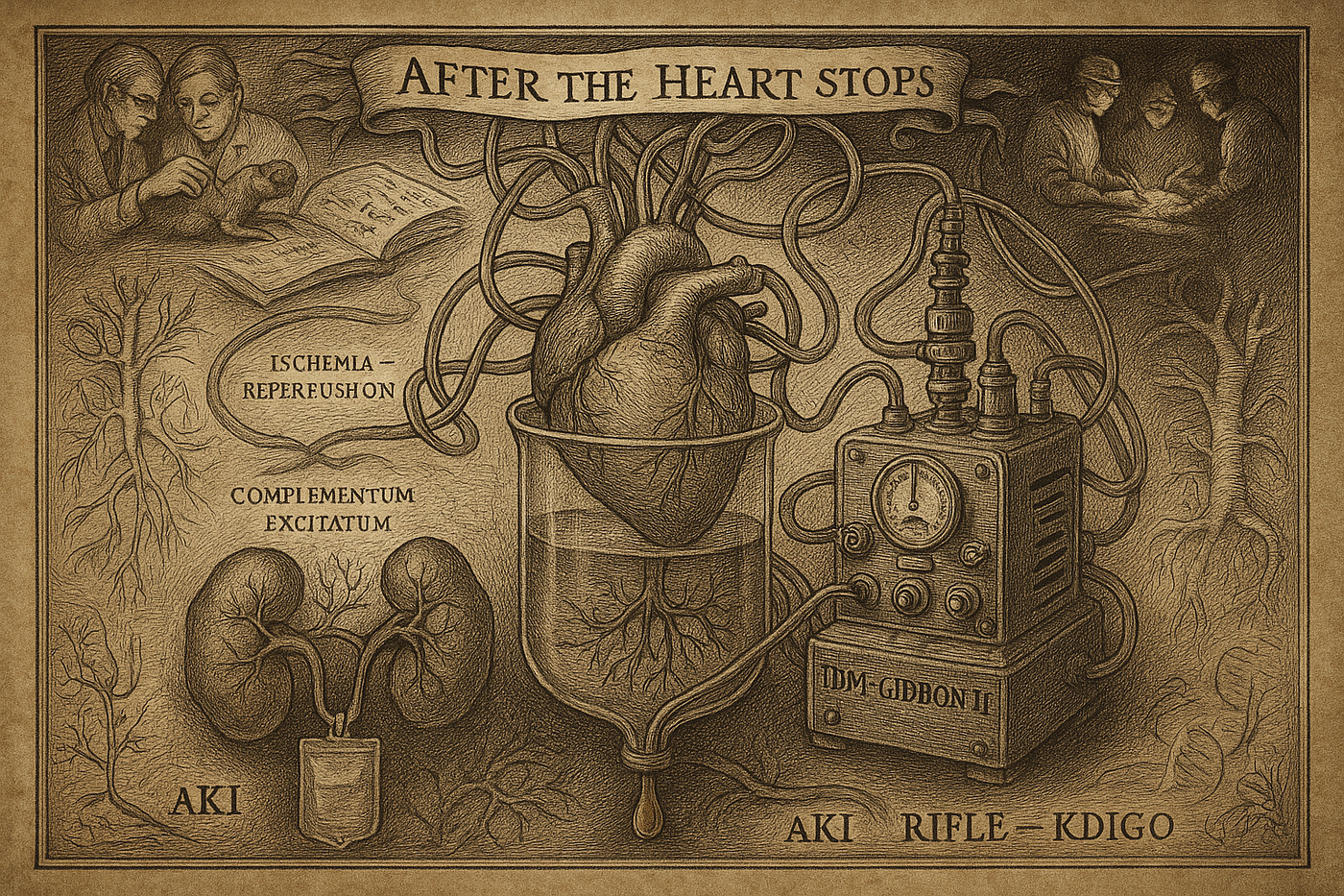
War interrupted. After it, fortune sent an IBM chairman across Gibbon’s path. The garage rig became the polished “IBM-Gibbon II.” Animal experiments mounted. Then reality intruded. His first human case, an infant, died on the table; the defect was more complex than anyone realized. The blow was crushing.
One year later, on May 6, 1953, an eighteen-year-old college student with an atrial septal defect lay on a table, connected to a gleaming tower of rollers and oxygenators. For twenty-six minutes a machine did her heart’s job while Gibbon stitched closed the hole. When her own heart took over and the blood turned cherry again, the room erupted. Humanity had crossed a line. Surgery inside a nonbeating, bloodless heart was no longer fiction.
Then two failures followed. The pioneer who labored twenty years to open a door into the heart never walked through it again. He left that dangerous and hope-filled space to those who came after. He could not have known that along with healing, he had released a ghost that still haunts our ICUs.
Pumphead, and a kidney that goes quiet
By the late 1950s the world had heard the thunderclap. Open-heart surgery spread quickly. Surgeons could finally work in a still, dry field, like watchmakers repairing a jeweled movement. Once the first exhilaration faded, strange patterns came into view.
The earliest whispers came from the brain. Families said their loved one was not quite themselves. Patients felt dulled, forgetful, less quick. We gave this constellation a blunt nickname, pumphead, or postperfusion syndrome. It taught us an uncomfortable truth. A heart-lung machine is a lifesaver, yet it is not organic blood flow. Microbubbles, tiny clots, and inflammation can leave subtle marks on the mind.
The kidney’s protest was quiet and more lethal. In the ICU, urine slowed to a trickle, then stopped. Urea and creatinine, nitrogenous wastes that healthy kidneys clear without fanfare, rose like a spring flood. Studies in the 1960s and 1970s made it plain. AKI after open-heart surgery was common, and when severe it carried a mortality in the range of two patients out of three despite dialysis. Surgeons could return a heart with triumph, only to watch a patient die days later from a kidney that would not recover.
The early explanation was mechanical. Longer time on bypass, lower pressures during surgery, weaker cardiac output afterward, all meant less perfusion. Starve a pump of water, the roof tank runs dry. That intuition contained a piece of truth, yet missed the scope of the storm. The heart-lung machine is not only plumbing. It stirs a biochemical weather system, fiercest in organs like the kidney where tiny vessels and delicate tubules sit at the edge between flow and hypoxia.
Name the enemy, see it clearly
For decades we could not even agree on what counted as AKI. More than thirty definitions littered the literature. A jump of 0.5 mg/dL in creatinine meant “failure” in one city, 1.0 in another, and “call me when they need dialysis” in a third. Studies could not talk to each other. We were in a Babel of our own making.
The turn came when nephrologists and intensivists gathered in Vicenza, Italy, and forged a common language. The 2004 RIFLE system reframed AKI as a spectrum that could be staged by simple parameters everyone owns, serum creatinine and urine output. The AKIN refinement in 2007 made the net tighter, recognizing that even a small bump in creatinine over forty-eight hours carries real risk. In 2012 KDIGO harmonized the two into the standard we use today. This was more than semantics. With a shared map, we could measure the true burden of CSA-AKI, compare strategies, and build trials that rhyme rather than argue. Naming the enemy was the first step toward defeating it.
What happens inside the kidney
Once you can see the target, you can visit the crime scene. CSA-AKI is not a simple robbery. It is a coordinated attack.
The opening blow is ischemia–reperfusion injury. During bypass, flow shifts from the heart’s pulsatile beat to the pump’s nonpulsatile stream. Pressures may sag. The kidney lives in relative drought. When the heart restarts, blood rushes back like a sudden storm. Oxygen sparks oxidative stress, spawning free radicals that batter cell membranes. The proximal tubules, workhorses of reabsorption, take the brunt, which is why acute tubular necrosis is the classic lesion after cardiac surgery.
Then friendly fire lands. Blood sliding across artificial tubing wakes the complement system, platelets, and white cells. Cytokines bloom. The intent is defense; the effect is a systemic inflammatory response that increases endothelial leak, clogs microcirculation, and deepens hypoxia within the kidney’s fragile cortex and medulla.
Accomplices join. Roller pumps shear red cells and spill hemoglobin into plasma. Free hemoglobin is directly toxic to tubules and can precipitate within them like silt choking a drain. Microemboli of air, fat, and platelet aggregates pepper the smallest vessels. On top of this, nephrotoxic exposures accumulate from contrast dye to broad-spectrum antibiotics to nonsteroidal analgesics. In a healthy organ each insult might pass. In a kidney under siege, the last straw breaks early.
This complexity explains why silver bullets have failed. A single antidote cannot block every doorway. We need systems thinking, prevention across the arc of care, and early warnings that buy time.
From firefighting to fire prevention
For years our stance was reactive. Watch creatinine and urine. When they drifted the wrong way, act and hope. Often that meant supportive care and, if the slide continued, dialysis. It was too late to rewrite the story.
Now the center of gravity has shifted toward prevention, a care bundle that starts in clinic and runs through the ICU.
Before surgery: know the terrain. Risk is not evenly distributed. Older age, female sex, reduced ejection fraction, diabetes, and especially preexisting chronic kidney disease raise the stakes. Optimize where you can: tune glycemic control, address anemia, prune medications that might amplify injury under stress. Temporarily hold agents like ACE inhibitors or ARBs twenty-four to forty-eight hours preop if the hemodynamic plan suggests vulnerability.
In the OR: make delivery the goal. Perfusionists and anesthesiologists have moved beyond “acceptable blood pressure” to goal-directed perfusion, targeting oxygen delivery to tissues, often cited around 270 mL per minute per square meter or higher. Pressure is a proxy; delivery is the payload. Temperature management matters, too. Avoid hyperthermia during bypass, which can worsen organ injury. Use balanced crystalloids rather than high-chloride saline to avoid renal vasoconstriction. Where it fits, techniques that reduce the physiologic insult, whether off-pump coronary bypass in suitable patients or biocompatible coatings on bypass circuits, can shrink the inflammatory footprint.
After surgery: anticipate, detect early, intervene early. In the ICU we watch pressure, output, cardiac index, and blood glucose with distrustful care, and we minimize nephrotoxic exposures. The weak link here is time. Creatinine is a rear-view mirror. By the time it rises, the crash already happened.
Which is why the search for early biomarkers matters. Molecules such as NGAL, or the product of TIMP-2 and IGFBP7, surge when tubular cells are stressed, often a day or two before creatinine whispers trouble. That lead time can be the difference between turning down a harmful vasoconstrictor, adjusting fluids with precision, or holding a drug that would otherwise tip the balance.
None of these steps is flashy. They are small, careful choices aligned toward a single aim: keep the kidney perfused, uninflamed, and unpoisoned while the heart is healed.
The weight of a bag of urine
Return to the operating room at the start. This time the patient rolls into the ICU, and the nurse’s first words are simple. “Eighty milliliters in the first hour.” No applause. No cheers. Yet those quiet numbers are more comforting than any postoperative chest film. A warm, golden bag of urine is not a given. It is the distilled outcome of seventy years of hard lessons. It holds Gibbon’s long night with a dying patient, the early surgeons’ defeats in the ICU, the scientists who mapped a messy physiology, and today’s teams who fuss over temperatures, pumps, fluids, and medicines as if they were jazz musicians tuning a standard.
The struggle between heart surgery and kidney survival may never vanish. As long as we must still the heart to mend it, tension will exist. What has changed is our frame. This is not a zero-sum game. We can protect a tired pair of kidneys while saving a failing heart. The journey from heroic feats to disciplined systems is the shape medical progress often takes. It is less cinematic than a single cure. It is more faithful to how people get better.
Keywords: cardiac surgery; acute kidney injury; ICU medicine; kidney protection; medical history
References
- Gibbon JH Jr. Application of a mechanical heart and lung apparatus to cardiac surgery. Minn Med. 1954;37(3):171–85.
- Romaine-Davis A. John Gibbon and His Heart-Lung Machine. Philadelphia: University of Pennsylvania Press; 1992.
- Abel RM, Buckley MJ, Austen WG, Barnett GO, Beck CH Jr, Fischer JE. Renal failure after open heart surgery. Ann Intern Med. 1976;84(6):677–82.
- Doberneck RC, Reiser MP, Lillehei CW. Acute renal failure after open-heart surgery utilizing extracorporeal circulation and total body perfusion. Analysis of 1000 patients. J Thorac Cardiovasc Surg. 1962;43:441–52.
- Bellomo R, Ronco C, Kellum JA, Mehta RL, Palevsky P; Acute Dialysis Quality Initiative workgroup. Acute renal failure — definition, outcome measures, animal models, fluid therapy and information technology needs: the Second International Consensus Conference of the Acute Dialysis Quality Initiative (ADQI) Group. Crit Care. 2004;8(4):R204–12.
- Mehta RL, Kellum JA, Shah SV, Molitoris BA, Ronco C, Warnock DG, et al.; Acute Kidney Injury Network. Acute Kidney Injury Network: report of an initiative to improve outcomes in acute kidney injury. Crit Care. 2007;11(2):R31.
- Kidney Disease: Improving Global Outcomes (KDIGO) Acute Kidney Injury Work Group. KDIGO Clinical Practice Guideline for Acute Kidney Injury. Kidney Int Suppl. 2012;2(1):1–138.
- Wang Y, Bellomo R. Cardiac surgery-associated acute kidney injury: risk factors, pathophysiology and treatment. Nat Rev Nephrol. 2017;13(11):697–711.
- Rosner MH, Okusa MD. Acute kidney injury associated with cardiac surgery. Clin J Am Soc Nephrol. 2006;1(1):19–32.
- Thakar CV, Arrigain S, Worley S, Yared JP, Paganini EP. A clinical score to predict acute renal failure after cardiac surgery. J Am Soc Nephrol. 2005;16(1):162–8.
- Brown JR, Kramer RS, Coca SG, et al. The Society of Thoracic Surgeons/Society of Cardiovascular Anesthesiologists/American Society of Extracorporeal Technology Clinical Practice Guidelines for the Prevention of Adult Cardiac Surgery-Associated Acute Kidney Injury. Ann Thorac Surg. 2023;115(1):34–42.
- Haase M, Bellomo R, Devarajan P, Schlattmann P, Haase-Fielitz A; NGAL Meta-analysis Investigator Group. Accuracy of neutrophil gelatinase-associated lipocalin (NGAL) in diagnosis and prognosis in acute kidney injury: a systematic review and meta-analysis. Am J Kidney Dis. 2009;54(6):1012–24.
- Kashani K, Al-Khafaji A, Ardiles T, et al. Discovery and validation of cell cycle arrest biomarkers in human acute kidney injury. Crit Care. 2013;17(1):R25.
- Newman MF, Kirchner JL, Phillips-Bute B, et al. Longitudinal assessment of neurocognitive function after coronary-artery bypass surgery. N Engl J Med. 2001;344(6):395–402.
- Lassnigg A, Schmidlin D, Mouhieddine M, et al. Minimal changes of serum creatinine predict prognosis in patients after cardiothoracic surgery: a prospective cohort study. J Am Soc Nephrol. 2004;15(6):1597–605.
- Zarbock A, Kellum JA, Schmidt C, et al. Effect of Early vs Delayed Initiation of Renal Replacement Therapy on Mortality in Critically Ill Patients With Acute Kidney Injury: The ELAIN Randomized Clinical Trial. JAMA. 2016;315(20):2190–9.
- Goren O, Matot I. Perioperative acute kidney injury. Br J Anaesth. 2015;115 Suppl 2:ii3–14.
메타데이터
- post_id
- 916d2f64159f
- slug
- after-the-heart-stops-916d2f64159f
- url
- https://medium.com/medical-stories/after-the-heart-stops-916d2f64159f
- canonical_url
- https://medium.com/medical-stories/after-the-heart-stops-916d2f64159f
- author_url
- https://medium.com/@critnephro
- status
- ok
- fetched_at
- 2026-06-09 15:37:30