The Metabolic Corridor: How the Gut’s Ecological Balance Shapes the Chemistry of the…
The physical distance between the colon, the liver, and the brain is large, yet the chemical traffic between them is continuous and tightly…
The Metabolic Corridor: How the Gut’s Ecological Balance Shapes the Chemistry of the Gut–Liver–Brain Axis
The physical distance between the colon, the liver, and the brain is large, yet the chemical traffic between them is continuous and tightly regulated. For decades this corridor was described in relatively simple terms. The gut was treated either as a passive reservoir of inflammatory triggers such as lipopolysaccharide or as a fermentation chamber that released short-chain fatty acids serving mainly as generic energy substrates [1,2]. Both framings missed the spatial and chemical logic that links the three organs. The gastrointestinal tract is now understood as a spatially ordered ecological system whose metabolic outputs are selectively filtered, transformed, and delivered to distant organs in distinct chemical languages [3,4].
Those languages — bacterial sphingolipids, host–microbe tricarboxylic-acid-cycle intermediates, and neuroactive tryptophan catabolites — do not act as diffuse signals [3,5]. They exert precise transcriptional, structural, and receptor-mediated control over insulin sensitivity, hepatic lipid handling, and neuroimmune tone [3,5,6]. Three principles emerge from this evidence. First, the chemistry that reaches the liver and brain is the residue of an ecological competition, not a portrait of the community that produces it [7]. Second, physical gradients of oxygen and mucus structure that competition [8,9]. Third, the same molecular families that drive hepatic lipid remodeling also set neuroimmune thresholds, so interventions at the level of the gut reverberate across the entire corridor [5,6]. Understanding the corridor therefore begins with the ecology that generates the chemistry.
Two Competing Guilds and the Logic of Relational Stability
The question that organizes this section is straightforward: why categorize the microbiota functionally rather than taxonomically? Because individual species are unstable units of prediction; functional networks survive where species do not. Contemporary systems approaches therefore organize the colonic microbiota into two self-reinforcing functional networks that compete for space and resources [7]. These networks are usefully termed the C1A Foundation Guild and the C1B Pathobiont Guild [7].
The C1A guild comprises strict obligate anaerobes — exemplified by Faecalibacterium prausnitzii, Roseburia intestinalis, Eubacterium rectale, and Ruminococcus bromii — that specialize in the hydrolysis of complex plant polysaccharides, or microbiota-accessible carbohydrates [2,10]. Their enzymatic breadth sustains cleavage of β-glycans that the host genome cannot process, and the products feed the colon [2]. Their principal products are the short-chain fatty acids acetate, propionate, and butyrate. Butyrate in particular serves as the physiological cornerstone of mucosal homeostasis and a tolerogenic immune tone [2,10].
The C1B guild consists of facultative anaerobes and opportunistic organisms, including members of the Enterobacteriaceae, Enterococcus, Fusobacterium, and certain Bacteroides [4,9]. Their genomes are enriched for virulence factors, antibiotic-resistance genes, and mucin-degrading enzymes. Under healthy conditions these organisms remain numerically and spatially constrained; when dietary or immunological barriers erode, they expand and drive both local and systemic inflammation [4,9].
The contrast is not taxonomic; it is ecological. Each guild deploys a different metabolic strategy against the same substrate, and the host physiology is the loser if C1B wins.
Earlier attempts to define a “core” human microbiome relied on taxonomic prevalence thresholds. That approach produced inconsistent results because low-abundance keystone taxa were frequently omitted and because strains within the same species can behave very differently [11,12]. The competing-guilds framework replaces prevalence with relational stability: the consistent co-variation and ecological coupling of specific genomes across dietary shifts, physical perturbations, and disease states [7]. Functional networks, not arbitrary taxonomic lists, become the unit of prediction.
Dietary fiber is the primary exogenous driver of this equilibrium [4,13]. When microbiota-accessible carbohydrates are abundant, the extensive carbohydrate-active enzyme repertoires of C1A organisms allow them to dominate. Prolonged fiber deprivation progressively depletes these taxa; in multi-generational animal experiments the loss can become irreversible [4]. Simply restoring fiber is then insufficient; the missing organisms themselves must be reintroduced, for example through fecal microbiota transplantation or carefully designed synbiotics [4,13].
Oxygen, Mucus, and the Physical Arena of Competition
Guild identity alone does not determine outcome; physical gradients do. The competitive balance is mapped onto the radial architecture of the colon.
Oxygen from the epithelial capillary bed diffuses inward and creates a steep gradient that is highest adjacent to the mucosa and lowest in the lumen [8,14]. Facultative C1B organisms colonize the oxygenated niche closest to the epithelium; obligate C1A organisms are confined to the hypoxic lumen. This segregation is reinforced by host metabolism rather than passively inherited from diffusion per se. Luminal butyrate drives β-oxidation in colonocytes, consuming the inward-diffusing oxygen and sustaining the anaerobic sink that excludes pathobionts from the privileged niche [8,14]. C1A metabolism therefore does double duty: it feeds the colonocyte and physically displaces the competitor.
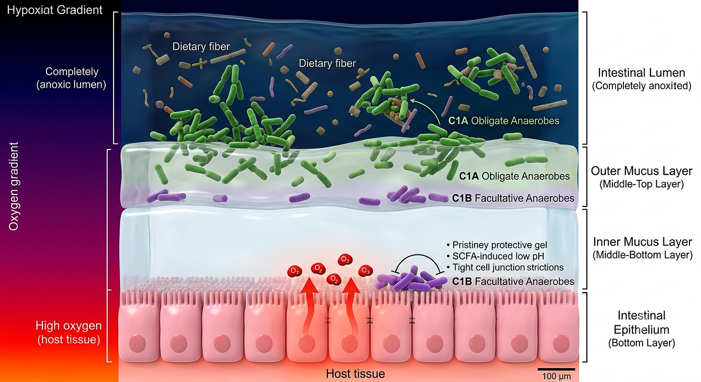
Fig. 1. Physical Arena of Competition. Schematic showing the intestinal lumen outer mucus layer, inner mucus layer and the intestinal epithelium with oxygen diffusion gradient. Due to differences in dietary fiber and the influence of oxygen spatial gradient, the microbiota evolves into two mutually competitive guilds, resulting in different spatial distributions.
A double-layered MUC2 mucus barrier further structures the space. The inner layer remains essentially sterile; the outer layer functions as a habitat and cross-feeding hub [9,15]. Specialized mucin degraders such as Akkermansia muciniphila release acetate and oligosaccharides that feed neighboring butyrate producers, sustaining a basal level of barrier support even during periods of low fiber intake [15,16]. When fiber deprivation is prolonged, the entire community shifts toward host glycan consumption. The protective inner mucus thins, C1B organisms establish biofilms on the epithelial surface, and inflammatory cascades are triggered [4,9]. The ratio of microbial genes encoding mucin-degrading versus plant-degrading carbohydrate-active enzymes — the MUC2plant ratio — provides a quantitative signature of this transition [4]. Tracking MUC2plant across cohorts offers a way to monitor functional collapse before clinical disease manifests.
Portal Filtration and Parallel Chemical Languages
The chemical output of this ecology reaches the liver through a single bottleneck: the portal vein. Every molecule absorbed from the gut must pass through the portal circulation. The liver therefore functions as both metabolic filter and primary recipient of gut-derived signals [3,5]. Metabolomic surveys show that portal blood is enriched for more than a hundred unique metabolites relative to peripheral circulation under standard diets; high-fat feeding sharply reduces this diversity [5]. A notable exception is a family of five-carbon dicarboxylic acids — aconitate, mesaconate, and 2-hydroxyglutarate — that remain consistently enriched regardless of diet, marking them as stable chemical links between gut activity and hepatic function [5].
The signals that dominate the dialogue with the liver travel in structurally unrelated chemical languages.

Fig. 2. Portal-Vein Corridor Function. Schematic of the anatomical pathway and functional role of the portal vein. All gut-absorbed metabolites must traverse the portal circulation before reaching the systemic compartment. The liver therefore acts as both metabolic filter and primary recipient of bacterial sphingolipids, host–microbe C5 dicarboxylates, and other ecological signals that rewire hepatic lipid handling and insulin sensitivity.
Bacterial sphingolipids. Members of the Bacteroidetes synthesize sphingolipids with saturated sphingoid backbones, odd-chain fatty acids, and distinctive head-group modifications [3]. Genetic ablation of serine palmitoyltransferase in Bacteroides thetaiotaomicron demonstrates that these lipids cross the epithelium, enter portal blood, and are incorporated into host metabolic pathways [3]. In the liver they remodel ceramide pools, alter de novo sphingolipid synthesis, and improve insulin sensitivity. Colonization of germ-free mice with wild-type B. thetaiotaomicron increases hepatic ceramides and reverses steatosis in experimental models; the corresponding SPT-deficient strain produces the opposite phenotype [3]. Bacterial sphingolipids therefore act as endogenous structural signals that rewire host lipid metabolism.
C5 dicarboxylates. Operating in parallel is a transcriptional network driven by itaconate and its isomers mesaconate and citraconate [5]. Itaconate is classically produced by activated macrophages via cis-aconitate decarboxylase (ACOD1/IRG1). Once released, it undergoes systemic turnover; roughly 5 % is converted to mesaconate, with the liver and kidney as major sites of interconversion [5]. Citraconate, generated along the same routes, acts as an endogenous inhibitor of ACOD1, providing negative feedback [5].
Portal concentrations of these dicarboxylates are elevated by approximately 1.5-fold after vancomycin disruption of the microbiota and correlate with the abundance of Akkermansia, Proteus, and Lactobacillales [5]. Both microbial and host TCA-cycle enzymes are upregulated, indicating mixed host–microbe origin. At physiological portal concentrations (~10 µM) the compounds increase insulin-receptor and Akt phosphorylation in hepatocytes, suppress lipogenic genes (SREBP1C, ACC1, FAS, SCD1, DGAT1) by 25–76 %, and elevate fatty-acid oxidation genes (PGC1α, PPARα, ACOX1, ACSL1, MCAD) two- to threefold [5]. These effects are PPARα-dependent. In high-fat-diet mice the same molecules restore insulin sensitivity to levels seen in lean controls. Human data from the Jackson Heart Study show an inverse correlation between circulating citraconate and fasting glucose, suggesting conservation across species [5].
The sphingolipid and C5 pathways diverge in chemistry but converge in effector logic: both rewire hepatic lipid handling through PPARα- and ceramide-dependent programs. Gut–liver signaling is therefore not a single circuit but an ensemble of structurally unrelated languages read by overlapping hepatic targets.
Extension to the Central Nervous System
The same chemical languages reach past the liver. Short-chain fatty acids and tryptophan catabolites reach the brain both by direct circulation across the blood–brain barrier and via vagal afferents [6,17].
Butyrate and propionate engage free-fatty-acid receptors (GPR41/43/109A) on enteroendocrine, immune, and endothelial cells, stimulating gut-hormone release, inhibiting histone deacetylases, and suppressing NF-κB-driven inflammation [2,6]. In the central nervous system they help maintain microglia in a homeostatic rather than reactive state, thereby moderating the neuroinflammation associated with Alzheimer disease, Parkinson disease, and amyotrophic lateral sclerosis [6,17].

Fig. 3. Pathways of Host-Microbiota-Derived Metabolites. Schematic summary of the principal chemical languages that travel the gut–liver–brain corridor. Bacterial sphingolipids and C5 dicarboxylates predominantly rewire hepatic lipid metabolism and insulin sensitivity, while short-chain fatty acids and tryptophan catabolites modulate microglial state, AhR signaling, and neuroimmune tone. All pathways remain under the upstream control of the C1A–C1B ecological equilibrium.
Microbial tryptophan metabolites — indole-3-propionic acid, indole-3-acetic acid, tryptamine — act as ligands for the aryl hydrocarbon receptor [6,18]. AhR activation promotes IL-22 production, strengthens tight junctions, and, within the brain, dampens pro-inflammatory programs in astrocytes [6,18]. Under dysbiotic conditions, however, inflammatory induction of indoleamine 2,3-dioxygenase shunts tryptophan toward the kynurenine pathway and the neurotoxic metabolite quinolinic acid, promoting excitotoxicity and protein aggregation [6,18]. Preservation of the C1A guild keeps this shunt in check.
In the liver, the relevant currencies are lipids and insulin-sensitivity programs; in the brain, the currencies shift to microglial state and neurotransmitter precursor balance. The chemistry differs, but the upstream controller is the same: the C1A–C1B equilibrium. This is the corridor’s load-bearing generalization.
Limits of Translation
The evidence above was produced largely in cell culture and rodent models, where spatial organization, microbiota composition, and metabolite flux differ from human physiology along three axes. Anatomically and ecologically, microbial colonization of intestinal crypts is pronounced in mice but largely restricted to the outer mucus layer in humans [8,14]. Kinetically, fluxes of short-chain fatty acids and tryptophan catabolites differ, so dose and kinetic parameters derived from animals cannot be assumed to transfer directly [6,17]. Pharmacologically, human trials must contend with genetic variation (for example, FUT2 secretor status), dietary background, and concurrent medications that reshape baseline microbiota and metabolite profiles [13].
Two further constraints make translation non-linear. The relationship between fiber intake and short-chain fatty acid production is saturating: moderate intake (20–30 g day⁻¹) yields metabolic benefit; higher amounts often produce only gas and bloating once the C1A metabolic machinery saturates [13]. And directionality is tissue-specific. While butyrate is anti-inflammatory in the colon, elevated systemic levels can drive pro-inflammatory Th17 responses in extra-intestinal tissues such as the kidney [19]. A metabolite that heals the mucosa can therefore inflame the kidney, depending on where it is read. The pharmacology of the corridor will therefore be a pharmacology of gradients, not a pharmacology of single molecules.
Closing Perspective
The gut–liver–brain axis is not a chain of organs connected by single molecules. It is a corridor whose output is the integrated product of an ecological competition, a physical gradient, and a series of distinct chemical languages read by different effectors in the liver and brain. Each step in the chain is dispensable when considered alone; the system depends on the relationships between them.
Three lessons follow. First, dysbiosis is not a single lesion but a failure of relational stability, and clinical trials that target individual species or metabolites will continue to underestimate this fragility [7]. Second, because the same molecular families govern hepatic lipid handling and microglial state, the corridor offers a rational target for interventions that act across organ boundaries — provided those interventions respect tissue-specific thresholds rather than assuming uniform benefit [5,6]. Third, the precision pharmacology the corridor demands cannot be borrowed from mouse models or from one-target-one-disease doctrine; it will require quantitative mapping of dose–response, saturation, and tissue-specific readout in humans.
The corridor is chemically precise. Navigating it will demand equal precision, and the first task is to measure relationships rather than chase single levers.
References
- Koh A, De Vadder F, Kovatcheva-Datchary P, Bäckhed F. From dietary fiber to host physiology: short-chain fatty acids as key bacterial metabolites. Cell. 2016;165(6):1332–1345.
- Louis P, Flint HJ. Formation of propionate and butyrate by the human colonic microbiota. Environ Microbiol. 2017;19(1):29–41.
- Johnson EL, Heaver SL, Waters JL, et al. Sphingolipids produced by gut bacteria enter host metabolic pathways impacting ceramide levels. Nat Commun. 2020;11(1):2471.
- Desai MS, Seekatz AM, Koropatkin NM, et al. A dietary fiber-deprived gut microbiota degrades the colonic mucus barrier and enhances pathogen susceptibility. Cell. 2016;167(5):1339–1353.e21.
- Portal vein-enriched metabolites as intermediate regulators of the gut microbiome in insulin resistance. Cell Metab. 2025. doi:10.1016/j.cmet.2025.07.014.
- Cryan JF, O’Riordan KJ, Cowan CSM, et al. The microbiota-gut-brain axis. Physiol Rev. 2019;99(4):1877–2013.
- Zhao L. Relational stability: a new strategy for defining the human core microbiome. Phenomics. 2025;5(1):14–17.
- Albenberg L, Esipova TV, Judge CP, et al. Correlation between intraluminal oxygen gradient and radial partitioning of intestinal microbiota in humans and mice. Gastroenterology. 2014;147(5):1055–1063.e8.
- Johansson MEV, Sjövall H, Hansson GC. The gastrointestinal mucus system in health and disease. Nat Rev Gastroenterol Hepatol. 2013;10(6):352–361.
- Morrison DJ, Preston T. Formation of short chain fatty acids by the gut microbiota and their impact on human metabolism. Gut Microbes. 2016;7(3):189–200.
- Turnbaugh PJ, Ley RE, Hamady M, Fraser-Liggett CM, Knight R, Gordon JI. The human microbiome project. Nature. 2007;449(7164):804–810.
- Shade A, Handelsman J. Beyond the Venn diagram: the hunt for a core microbiome. Environ Microbiol. 2012;14(1):4–12.
- Makki K, Deehan EC, Walter J, Bäckhed F. The impact of dietary fiber on gut microbiota in host health and disease. Cell Host Microbe. 2018;23(6):705–715.
- Donaldson GP, Lee SM, Mazmanian SK. Gut biogeography of the bacterial microbiota. Nat Rev Microbiol. 2016;14(1):20–32.
- Belzer C, Chia LW, Aalvink S, et al. Microbial metabolic networks at the mucus layer lead to diet-independent butyrate and vitamin B12 production by intestinal symbionts. mBio. 2017;8(5):e00770–17.
- Derrien M, Vaughan EE, Plugge CM, de Vos WM. Akkermansia muciniphila gen. nov., sp. nov., a human intestinal mucin-degrading bacterium. Int J Syst Evol Microbiol. 2004;54(Pt 5):1469–1476.
- Dalile B, Van Oudenhove L, Vervliet B, Verbeke K. The role of short-chain fatty acids in microbiota–gut–brain communication. Nat Rev Gastroenterol Hepatol. 2019;16(8):461–478.
- Rothhammer V, Quintana FJ. The aryl hydrocarbon receptor: an environmental sensor integrating immune responses in health and disease. Nat Rev Immunol. 2019;19(3):184–197.
- Park J, Kim M, Kang SG, et al. Short-chain fatty acids induce both effector and regulatory T cells by suppression of histone deacetylases and regulation of the mTOR–S6K pathway. Mucosal Immunol. 2015;8(1):80–93.
메타데이터
- post_id
- 98ddfc035745
- slug
- the-metabolic-corridor-how-the-guts-ecological-balance-shapes-the-chemistry-of-the-98ddfc035745
- url
- https://medium.com/@jungyhuang/the-metabolic-corridor-how-the-guts-ecological-balance-shapes-the-chemistry-of-the-98ddfc035745
- canonical_url
- https://medium.com/@jungyhuang/the-metabolic-corridor-how-the-guts-ecological-balance-shapes-the-chemistry-of-the-98ddfc035745
- author_url
- https://medium.com/@jungyhuang
- status
- ok
- fetched_at
- 2026-08-19 02:54:09