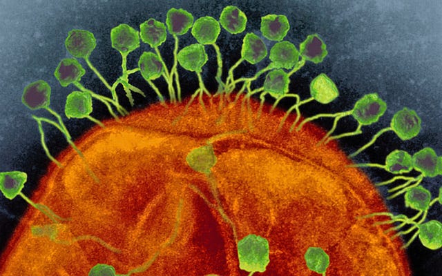
Methods and Applications for Genetically Engineering Phages to Further Phage Therapy Progress and…
Phage therapy, utilizing bacteriophages (phages), might be our last hope in combating antibiotic resistance. But, due to phage therapy…
Methods and Applications for Genetically Engineering Phages to Further Phage Therapy Progress and Combat Obstacles for Clinical Use
Abstract
Phage therapy, utilizing bacteriophages (phages), might be our last hope in combating antibiotic resistance. But, due to phage therapy being a biological treatment (dependent on living organisms), it is hard to ensure that the phages are consistent, successful, and safe to use each time it is administered. Because of this, phage therapy currently isn’t FDA-approved in Western countries, as well as many other countries around the world. Genetically engineering phages may be used to combat these obstacles and make phage therapy safe for clinical application. This paper covers six of the most common gene editing methods for phage engineering (homologous recombination, plasmid-based recombination, bacteriophage recombineering of electroporated DNA (BRED), PHEIGES, CRISPR-Cas systems, and adaptive evolution). Modifications that have taken place include reduction of genomes, improvement of phage stability in different environments, modified host receptors, mass-production of gene-edited phages, and new randomized insertions of genes.
Introduction
Antibiotic resistance has been developing at an alarming pace in bacteria due to the global overuse of antibiotics⁸. In 2019, it was estimated that antibiotic resistance was directly responsible for 1.27 million deaths and contributed to 4.95 million deaths. There has also been an increasing rise of bacteria that have evolved into superbugs, which are bacterial strains that are resistant to every antibiotic and other form of treatment we have. Because of this, those who develop infections from these superbugs have a greater risk of death compared to before, and there has already been a huge rise in deaths that are being reported⁸.
Phage therapy was first discovered in 1919 by the French-Canadian microbiologist Felix d’Herelle. It is a treatment utilizing naturally occurring bacteriophages (phages) to target specific bacteria that are causing bacterial infections in the body. It was abandoned after the discovery of penicillin, which led to the creation of antibiotics, and progress on phage therapy halted in most of the world. A few countries continued their research, and now, in the post-antibiotic era, with the rise of antibiotic-resistant bacteria leading to more deaths, more countries are taking an interest in this therapy. It is said that phage therapy is the final attempt to combat antibiotic resistance and that it is our only solution to prevent this global crisis from becoming a global problem.
Phages are very precise as to what bacterial strain they infect, so it makes them perfect for direct administration into the body. The phages are administered (can be through oral, injection, topical, aerosol, etc.) into the body and they will travel to locate the bacterial strain they need to target. Because of their specificity, it is very unlikely that they will interact with other components in the body while searching for the bacteria, minimizing any side effects or symptoms that arise with the use of antibiotics. Then, once the phages locate the target bacteria using receptors, which are unique molecules on the surface of a bacterial cell that phages recognize and bind to the phages, they can begin the infection process. Phages have two infection cycles, lytic and lysogenic, but for phage therapy, experts prefer using lytic phages. Lytic phages are more fast-acting by killing their bacterial host right away, which removes the chances of the target bacteria coming back. Lysogenic phages, on the other hand, put their bacterial host into “idle” mode after infecting it, and once that bacteria becomes active again, the lytic cycle will kickstart to finish the job.
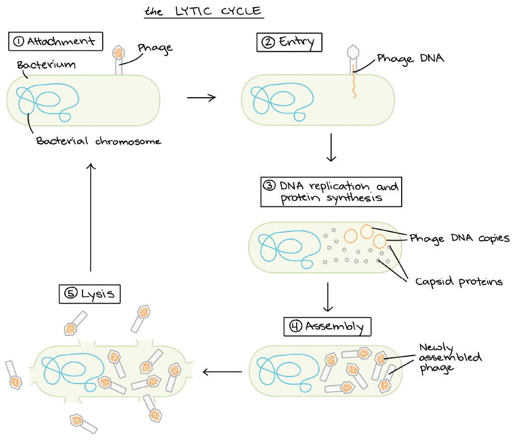
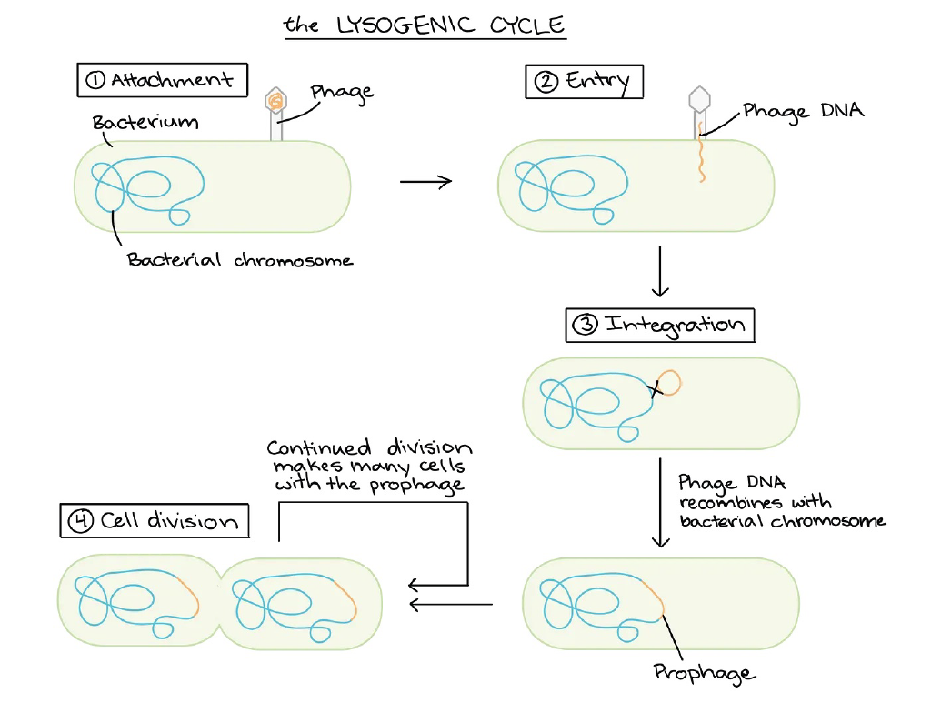
Left: A diagram of the lytic phage infection cycle. Right: A diagram of the lysogenic phage infection cycle. Photo credit
Focusing on the lytic cycle, the lytic phages then inject some of their own DNA into the bacteria. Then, the injected DNA begins a replication process, creating copied phage DNA and capsid proteins (the shell where the DNA is stored in a phage), which then causes the assembly of new phages to occur. These newly assembled phages then trigger lysis, which is where these phages basically penetrate and break out of the bacterial strain, causing it to burst and be destroyed, making it non-functional.
Although phage therapy has many positives for its use, many obstacles still need to be overcome. For example, because phages are so niche as to what they can infect, it can be difficult to find the specific phage species that will infect the exact bacterial strain we need to target. There are also concerns with phage stability in different temperature and pH environments, which can alter its effects when attacking bacteria, and the therapy can prove unsuccessful. Phages are also living organisms, so they can be unpredictable due to natural evolution when put in use for clinical applications. These are a few of the factors we need to take into consideration when deploying phage therapy, being the reason that it isn’t FDA-approved in most countries. Countries that have continued its development include Russia, Poland, and Georgia, which have pioneered phage therapy research and continue to use it regularly throughout those regions to this day.
Genetic engineering modifies an organism’s genome to enhance or remove traits, which can improve phage therapy. Utilizing different gene editing techniques on phages can reduce their genomes, which provides for more consistency on a larger scale and more predictability. Modifying phage receptor binding proteins can both change and increase a phage’s host range when determining the bacteria it can target, as well as possibly allowing it to target different diseases/conditions altogether that aren’t caused by bacteria, furthering phage therapy possibilities even more. Genetically engineering phage genomes could also improve phage stability in different environments for more scenarios and variability. Not only can genetic engineering of phage genomes be used to overcome these technical obstacles, but it can also be used to enhance the already good aspects of phage therapy to make it even better, like, for example, modifying the kind of “infecting substance” phages use (for example, instead of a phage injecting its DNA into a bacterial host when infecting it, it could possibly instead inject a drug or antibiotic).
Phage therapy has great potential, and genetically engineering phages can turn the potential into a reality. This paper will cover the different methods of genetically engineering phages as well as examples of how these methods can be applied and what kind of modifications can be made to phage genomes to improve their overall performance in phage therapy.
Methods for Phage Engineering
1. Homologous Recombination
Homologous recombination is one of the earliest techniques of genome editing. It is a naturally driven process that refers to the exchange of nucleotide sequences between two DNA molecules, which share similar or identical sequences¹. This process can be done by using two different (parental) phages that each have desirable traits and contain homologous regions in their DNA sequences. The parental phages co-infect a host bacterium and have their DNA injected. The infection of the bacterium then occurs and the phage DNA is replicated to create new phages. Because there were two parental phages co-infecting the bacterial host, both DNA sequences of those two phages can facilitate the exchange of genetic material and can create completely new phage progenies with traits from both parental phages. Then, lysis occurs and the phages are released¹.
In order to make specific modifications to phage genomes, and those traits aren’t occurring naturally in other species of phages, scientists have to integrate donor DNA templates that contain the desired gene edits¹. These templates need to have sequences of DNA that are homologous to the target regions in the phage genome. The homology in both DNA sequences acts as a guide to align the donor DNA with the target in the phage genome and can begin exchanging information between the two sequences to create the edits¹.
In order for this method to have success in editing the phage genome, it needs to be ensured that there is a sufficient amount of homologous region near the target site of the DNA sequence to promote the biological occurrence¹. This limits the potential for precise integration of the genetic changes in the genome, which is why homologous recombination is not widely used as a genome editing technique compared to other, more advanced options like BRED or CRISPR-Cas systems.
2. Plasmid-Based Recombination
Plasmid-based recombination is similar to homologous recombination engineering. It still requires sequences that are homologous to the target in the phage genome, but the designed mutation that is intended to be made is contained in a plasmid¹. The plasmid is constructed to contain the edits. Scientists have used molecular cloning to insert a gene of interest into the plasmid. The plasmid is then transformed into the chosen host bacteria through either transformation, transfection, or conjugation. The phage that is intended to be genetically modified then infects the bacteria with the plasmid. Then, the DNA begins to replicate and recombination can occur between the phage DNA and the donor template DNA that was contained in the plasmid. This then causes new phages with modified genomes to be made and released¹.
There are downfalls to plasmid-based recombination that are similar to homologous recombination, like requiring a sufficient amount of homologous DNA sequence in the plasmid as well as the limitations of how specific of a modification you can make. There are also issues with how accurate these modifications can be. Recombination, both homologous and plasmid-based, is not always accurate and unintended insertions or deletions may occur, which can lead to unwanted mutations. Besides some of those issues, plasmid-based recombination remains a valuable tool for creating genetic modifications in phages.
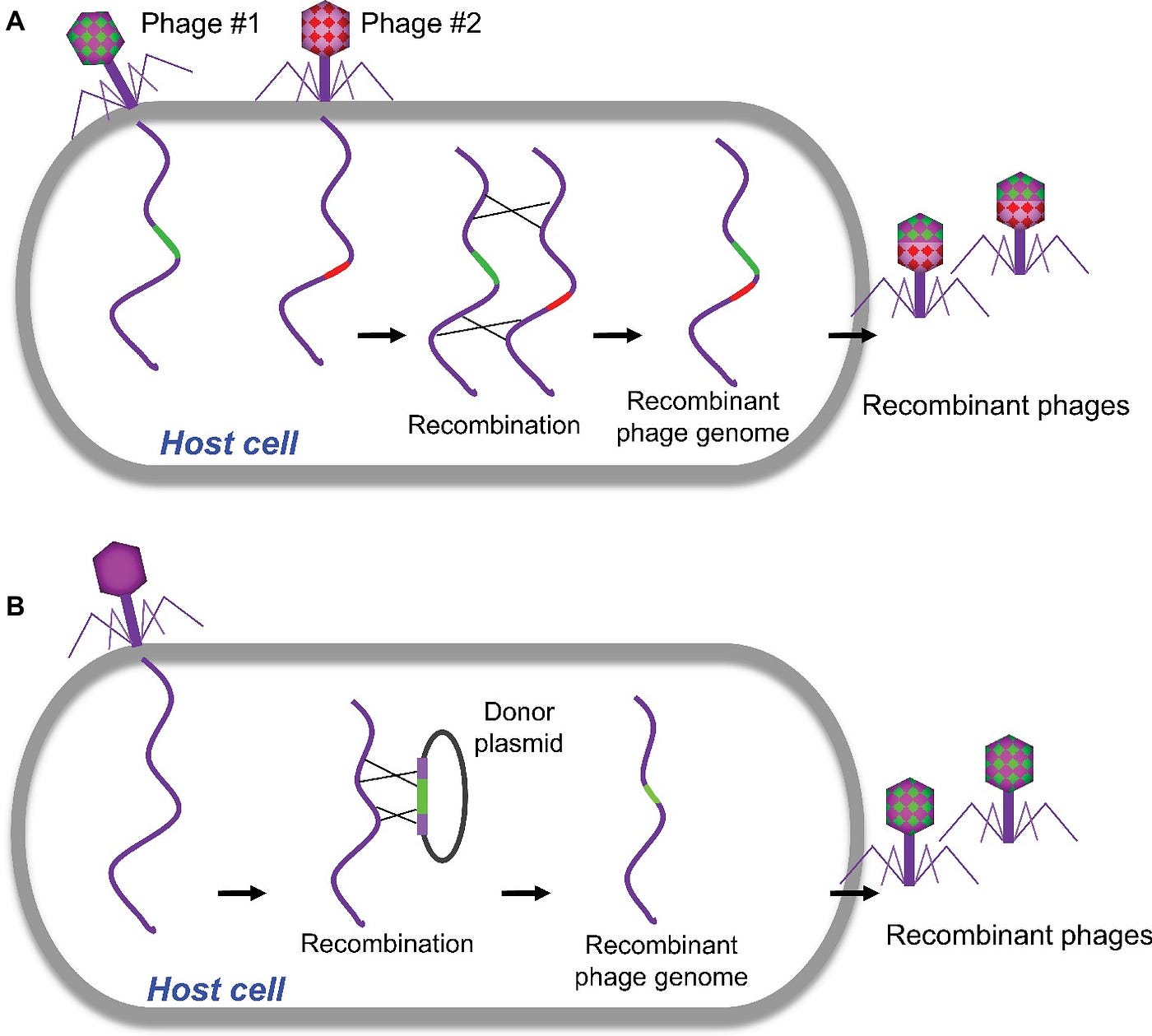
Two diagrams showing the differences and processes of homologous recombination and plasmid-based recombination. Photo credit
3. Bacteriophage Recombineering Of Electroporated DNA (BRED)
Bacteriophage Recombineering of Electroporated DNA (BRED) is a strategy used to edit a series of bacteriophage genes to improve the efficiency of the recombination approach. This gene-editing strategy takes place in a bacterial host¹. BRED uses a phage-encoded recombination system, for example, the Red system proteins of phage lambda or the RecE/RecT system proteins of the Rac prophage, to enhance the frequency of homologous recombination. If a phage doesn’t have encoded proteins such as these to promote homologous recombination, the system can be introduced to the bacterial host through plasmids or chromosomal insertions. Normally, this system is always found in bacteria, but the BRED technique requires either both the phage and the bacterial cell to have the homologous promoting proteins like RecE/RecT, or just requiring the bacterial cell to be genetically engineered further for enhancement of the system¹.
The process starts by using co-electroporation of the phage DNA template (which contains all the intended genetic modifications) and the donor DNA into the bacterial cells¹. Both of these DNA sequences also have homologous regions, which is required in any homologous-based recombineering, which BRED utilizes but in a more enhanced way. Then, the homologous recombination system activates and occurs between these two phage sequences, exchanging and rearranging the DNA sequences to produce new progeny phages with the desired mutations/traits. This method has limited use in Gram-positive bacteria, which would then exhibit low transformation efficiencies¹.
In an experiment conducted to modify the capsid proteins in E. Coli T7 phages to improve the resistance to acidic environments and digestive enzymes to improve the stability of orally injected phages for their use in veterinary medicine, the BRED method was used in making the modifications to the capsid⁷. The phage genome was modified using BRED to incorporate an E. Coli PhoE signal peptide into the major capsid protein to create T7::PhoE phages. T7L::PhoE phages were then exposed to conditions simulating the GIT, varying in temperature (30-42℃) and pH (2.5–5.7). The mutant phages demonstrated better survival under acidic conditions and resistance to digestive enzymes. Compared to wild-type T7 phages, phage T7::PhoE survived longer at pH 2.5 for up to 30 minutes compared to 15 minutes for wild-type phages. Higher titers were also retained for mutant phages compared to wild-type phages at higher temperatures and pH levels such as 3.5⁷.
While the BRED method worked for this experiment, in another paper, it was said that a single plaque obtained by using that method accounted for only 1% to 22% of the total plaques⁹.
BRED has become an important method for genetically engineering phages and has improved upon traditional methods such as homologous recombination and plasmid-based recombination.
4. PHEIGES: All-Cell-Free Phage Synthesis and Selection
PHage Engineering by In vitro Gene Expression and Selection (PHEIGES) is a novel method for speeding up the process of producing newly edited phage genomes, removing the need for a bacterial host. This both simplifies and accelerates the manufacturing of phages on a large scale. PHEIGES takes copies of already edited phage genomes and creates the building blocks for more phages to be created with that desired genome.
In a paper that utilized PHEIGES for T7 genome engineering, polymerase chain reaction (PCR) was used to amplify and create multiple copies of the desired phage genome to be coded into actual functional phages⁶. PCR was used because it is one of the few modern techniques that does not depend on a bacterial host, which can prove to be time-consuming for the process. After the DNA was amplified, it was assembled in vitro using a mix containing an exonuclease enzyme, which promotes the joining of all the new amplified fragments. The exonuclease enzyme was heat-inactivated to prevent it from modifying the newly constructed genome further. This resulted in multiple copies of DNA fragments that can be used as the template DNA for the phages⁶.
PHEIGES utilize an E. Coli cell-free transcription-translation (TXTL) system, which is a laboratory technique that offers experimental settings for the production of proteins without using living cells⁶. In the process, TXTL takes the DNA fragments (acting as template DNA) that are introduced and drives the production of phage proteins by carrying out transcription and translation (from DNA to RNA, then from RNA to protein) in vitro. The phage proteins from that process are then used to assemble functional phages with the desired genomes, directly from that one phage genome introduced in the TXTL reaction. In the paper that used PHEIGES to edit phage genomes, TXTL reactions were carried out in batch mode at scales of 1–10 μl, either in 1.5 ml tubes or in well plates. This process used in the paper had high phage yields, producing up to 10¹¹ plaque-forming units per millimetre (PFU/ml) of engineered phages within a day⁶.
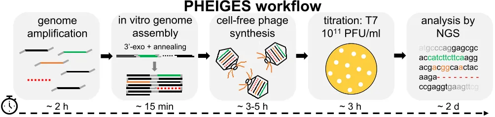
This is a flow chart of the PHEIGES process to create genetically engineered phages. Photo credit
In the paper, PHEIGES was used to create multiple copies of phages that had modified genomes encoded into them⁶. Three key phage genome engineering experiments tested the use of PHEIGES for reconstructing the genome edits into new phages. The first experiment was constructing phages with a genome that had an insertion of a mCherry fluorescent protein gene. The experiment was successful, the PCR process was able to amplify the segments of the DNA that were edited with the mCherry gene and create the proteins that preserved the edit when being used to construct the new phages. The second experiment used PCR to only amplify specific segments of a T7 phage genome, resulting in a newly constructed DNA template with specific deletions. The deletions were evaluated by considering which class the potential target genes came from. There are 3 classes of phage genes. Class 1 genes are early genes responsible for the initial stages of phage infection, class 2 genes are involved in DNA metabolism and replication (not strictly essential for phage survival), and class 3 genes are essential genes that are crucial for phage replication and infectivity. The T7 genome was divided into 8 fragments that had six targeted deletions of genes from classes 1 and 2. Deletions 2–4 were successfully made, while deletions 1, 5, and 6 did not result in viable phages. The T7 genome was originally 40.7 kbp (kilobase pairs) long, and after the combined deletions, was shortened to 35.8 kbp long, creating phage T7 mini. Phage T7 mini was still able to infect its natural host E. Coli B. The third experiment was to create a phage variant library to test out the success of PHEIGES despite a great variability in the phage genomes. Using PCR mutagenesis, random mutations were introduced into specific regions of phage genomes. The method works by intentionally introducing errors during amplification to generate a diversity of sequences. One edit that was made was the introduction of a tail fibre gene. The TXTL system was still able to synthesize and create the proteins for all these newly edited phage genomes that would be constructed into functional phages. The results indicated success in creating a library of phage variants with diverse host ranges. The newly created phages were able to target a broader range of bacteria compared to the wild-type phage⁶.
5. CRISPR-Cas Methods
CRISPR-Cas genome editing methods are widely used and are one of the most common techniques for creating genome edits, especially for genetically engineering phage genomes. CRISPR-Cas systems contain 2 main components for gene editing, the Cas proteins and the guide RNA (gRNA)¹. If the goal is to create an insertion/replacement, then there is also usually a homologous template DNA sequence, which would also be considered a main component. All of these components are most commonly delivered through plasmids, which are very versatile. The gRNA is designed to match the DNA sequence in the target region of the genome and binds with the Cas protein, forming a complex. This complex then searches the genome for the DNA sequences that match the gRNA, and the gRNA guides the Cas protein to where the intended edits are going to be made. The gRNA can then recognize the sequence of the target DNA. For the cut to be made, a short DNA sequence located near the target DNA, called a protospacer adjacent motif (PAM), needs to be verified by the Cas protein to confirm that this is the correct target DNA and that it’s okay for it to cut. The Cas protein then makes a double-stranded break at the specific site. Then, if there was the intent to make an insertion/replacement, the DNA template would be used to repair the break. If not, the ends of the break will join together, deleting the original gene that was cut altogether. CRISPR-Cas systems have been used in multiple phage genome editing experiments, and it has proved to be efficient, cost-effective, and scalable. The CRISPR-Cas system can be classified into 6 types and 2 broad classes (class 1 and class 2), which are determined by phylogeny and activity mechanisms. Class 1 includes methods 1, 3, and 4, which employ effector complexes containing multiple Cas proteins. Class 2 with methods 2, 5, and 6, employ effector complexes containing a single Cas protein to cleave the target DNAs¹.
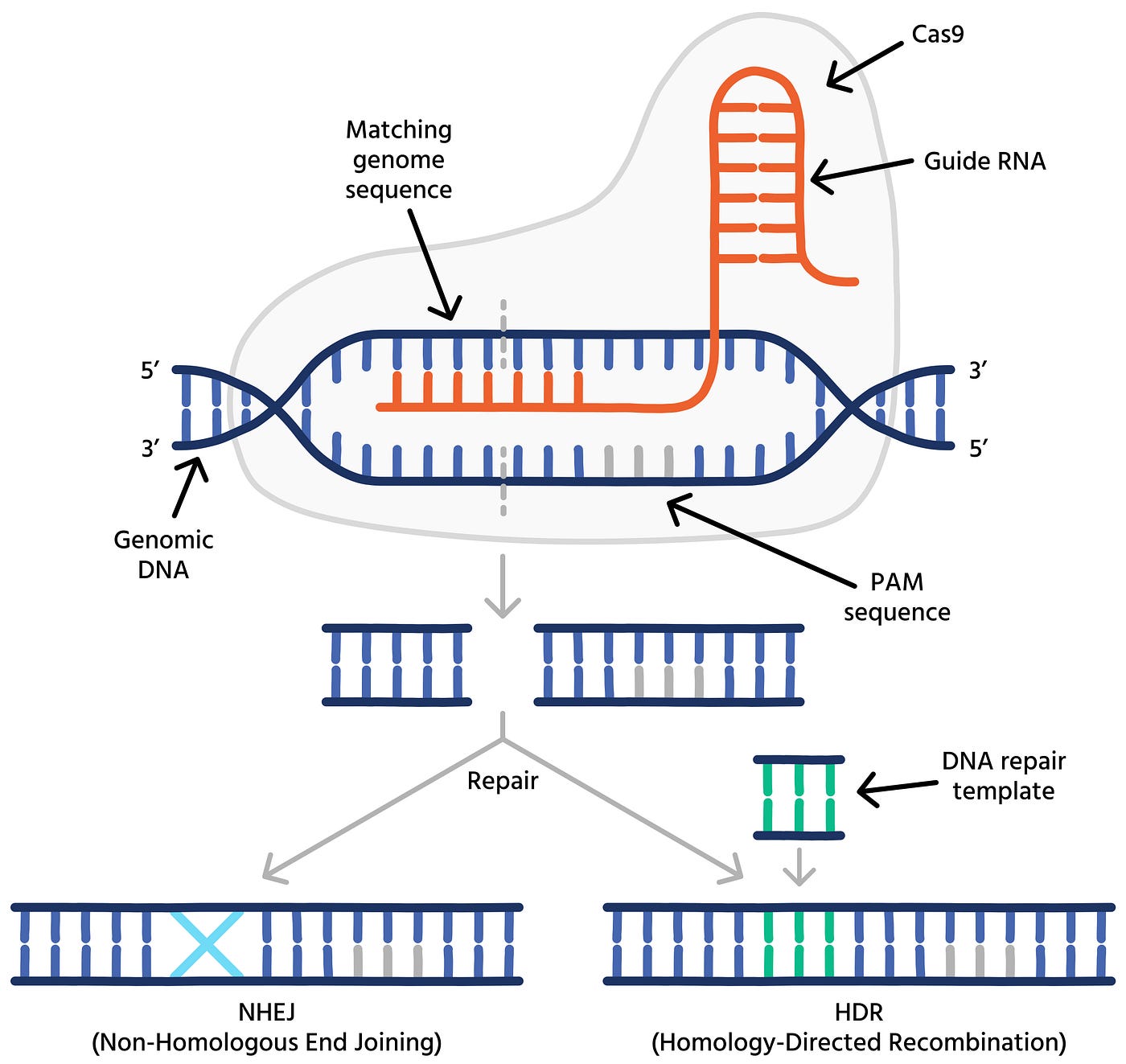
Diagram of CRISPR-Cas9 gene editing process in DNA strands. Photo credit
The CRISPR-Cas 9 complex is the most common and is considered to be the most versatile and efficient CRISPR-Cas system. It has been found that the Cas9 complex can efficiently cleave the T4 phage genome¹. In an experiment done to edit the genome of phage TT4P2, which infects Vibrio natriegens, CRISPR-Cas9-based systems were used⁹. The two CRISPR-Cas methods used were SPMAR (Single-Plasmid-Mediated Asynchronous Recombination), which utilizes a simpler, single-plasmid system. The other CRISPR-Cas method used was DPMSR (Dual-Plasmid-Mediated Synchronous Recombination), which used a more complex, dual-plasmid system. The experiment involved targeting two genes in the TT4P2 genome, gene orf6 (292 bp long) for deletion and replacement, and gene orf45 (162 bp long) for deletion. Gene orf6 was replaced with orf39 from Enterobacter myophage EJ9P3, which encoded lysozyme e, an enzyme that breaks down bacterial cell walls. For the edit of gene orf6, all the necessary components were stored and delivered in plasmid pT6ed, while for the edit of gene orf45, the components were stored and delivered in plasmid pT45ed. When the plasmid pT6ed was introduced to phage TT4::pT6sp2 (TT4P2 phage with inserted spacer 2), this had an increased EOP (efficiency of plating) for the phage. When the plasmid pT45ed was introduced to TT4::pT45sp1 (TT4P2 phage with inserted spacer 1), this had a decreased PFU by five orders of magnitude. These results indicated that the gene orf45 may be an essential gene for the phage’s life cycle. For the SPMAR method, 35 plaques were obtained and sequencing showed that 34/35 plaques were recombinants. The editing efficiency of the SPMAR method was 97%. For the DPMSR method, 19 plaques were obtained with all of them being recombinants. The editing efficiency of DPMSR was 100%. Some unwanted mutations occurred such as the entirety of orf6, the back of orf5 (121 bp) and the front of orf7 (182 bp). These results and unwanted deletions indicated that orf6, as well as orf5 and orf7, are non-essential genes that can be deleted without having an impact on the phage’s ability to produce. Both SPMAR and DPSMR methods successfully achieved high-efficiency genome editing in phage TT4P2⁹.

CRISPR-Cas9 gene editing method diagram for genetically engineering phages. Photo credit
In a different paper, the development and application of the CRISPR-cas12a system was explored for editing Pseudomonas aeruginosa phages and was compared to the widely used CRISPR-Cas9 system². The cleavage efficiency of both systems was tested for 3 P. aeruginosa phages (phage PJNP029, phage PJNP013, and phage PJNP053). For the cleavage efficiency test, 5-9 genome targets in each of the phages were randomly selected. The highest plaque formation efficiency from utilizing the Cas9 system in the phages’ edits was ~10⁴, while the highest plaque formation efficiency utilizing the cas12a system was ~10⁶, and in each test, the Cas12a system outperformed the Cas9 system with consistently higher cutting efficiency, ranging from 10 to 10⁴ times higher than Cas9. For editing the phage genomes, CRISPR-Cas12a was used for further testing of its abilities. For phage PJNP053, high and weak activity gRNAs were assembled with 500 bp homology arms into plasmids and were used with the Cas12a system to make deletions. With high-activity gRNA, the length of the deletion was 505 bp, while the length with the weak-activity gRNA was 538 bp. A gene insertion experiment was then conducted with phage PJNP029, where a 699 bp eGFP gene with 500 bp homology arms was inserted, and the Cas12a system achieved 100% positive clones. Lastly, homology arm testing was done with varying lengths from 30 bp-500 bp. Success rates ranged with lengths of 100 bp-500 bp, while no edits were observed with lengths of 30 bp-50 bp. Lastly, cross-species testing was done to see the effectiveness of the Cas12a system in different phages such as E. Coli phages T4 and T7, and Salmonella phages PJNS016 and vB_SalS_JNS02, which achieved 100% potentially positive clones and varying cutting efficiencies, with confirmed successful deletion or target genes. There was also genome reduction done in phages T7, T4, seszw, and selz phages, with reduced genome sizes by 8–23%. The CRISPR-Cas12a system was preferred compared to the more commonly used CRISPR-Cas9 due to its smaller protein size and easier-to-design RNA guide sequences, as well as it was more efficient compared to the Cas9 system².
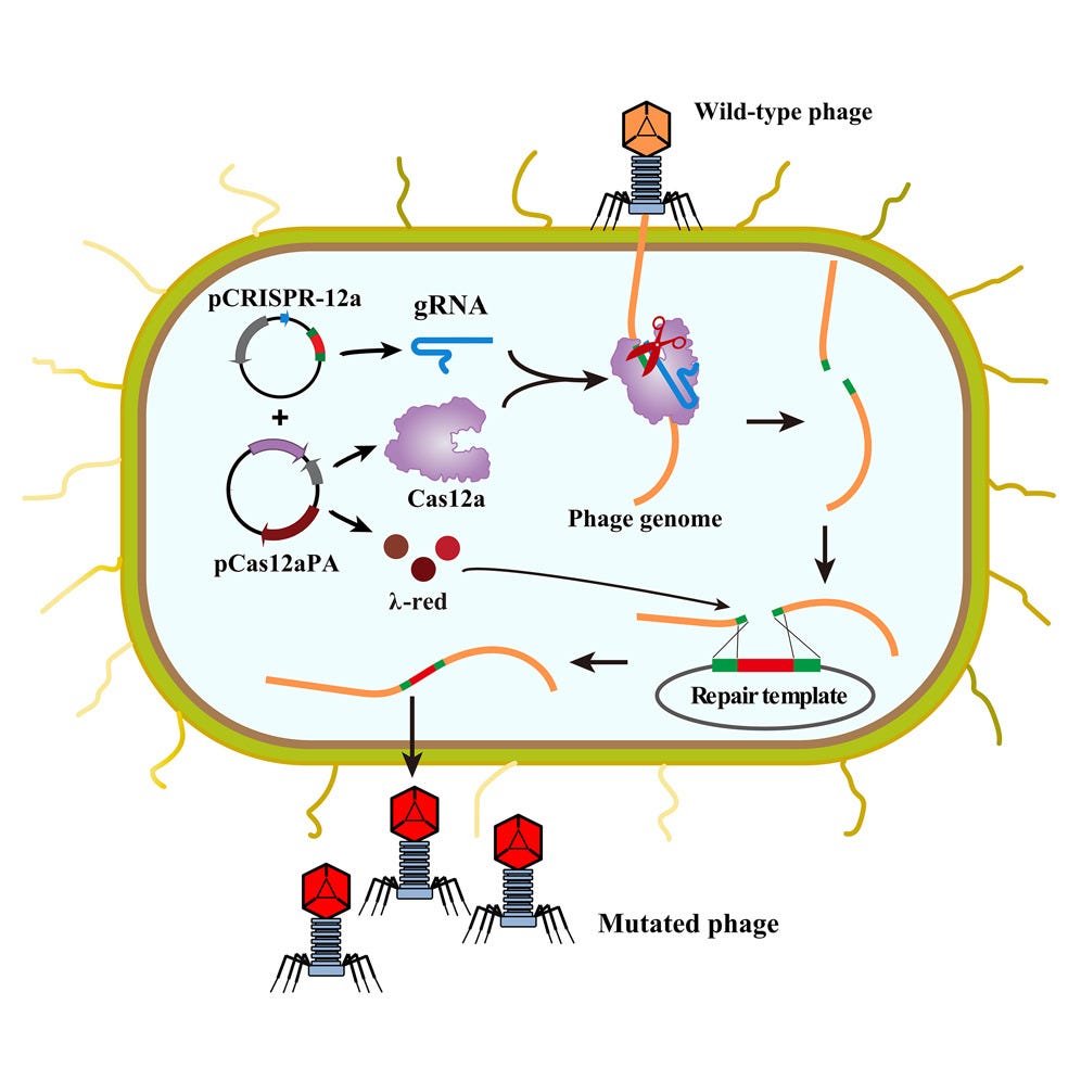
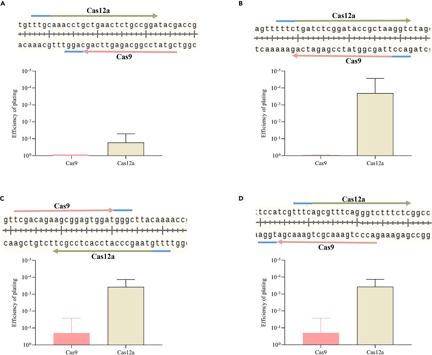
Left: A diagram of the CRISPR-Cas12a gene editing method. Right: Diagrams pulled from “Development of the CRISPR-Cas12a system for editing of Pseudomonas aeruginosa phages” comparing the results of utilizing Cas9 and Cas12 systems for CRISPR gene editing. Photo credit
Adaptive Evolution and Phage Stability
6. Using Adaptive Evolution as a Phage Engineering Method
While there are many techniques for genetically engineering phages, there are some that utilize the power of natural evolution and adaptation of species. In a paper focused on testing the stability of phages in different temperatures, they utilized adaptive evolution to enhance the phages’ stability in non-optimal conditions⁴. They were able to achieve this by exposing the phages to repeated cycles of heat stress. The goal was to develop phages with better resistance to storage conditions, especially at elevated temperatures, all while maintaining their ability to infect and destroy bacteria. The phages being subjected to the heat treatment were phage Wc4, phage CX5, and phage P-PSG-11⁴. In order to do this, the phages were exposed to cycles of heat treatment in a water bath at 60℃ for 1 hour, cooling at 28℃ for 15 minutes, overnight incubation on agar plates, centrifugation at 4℃ for 15 minutes to separate the phages, and repeated water bath exposure also at 60℃ for 1 hour. This process was repeated five times to encourage the phages to naturally adapt. This heat-treatment cycle created phage Wc4–1, phage CX5–1, and phage P-PSG-11–1⁴.
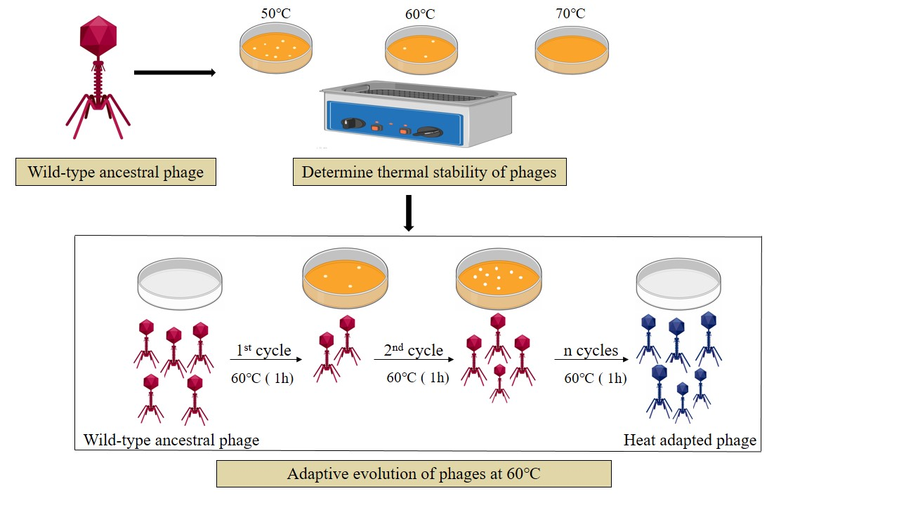
Graphic of methods for adapting the phages to a higher level of heat resistance as well as methods for testing resistance rates in both wild-type and ancestral phages. Photo credit
After this heat treatment was performed for all the phages, both ancestral and heat-treated phages were exposed to high temperatures for durations of 14, 30, and 60 days to test their stability⁴. The ancestral and heat-treated phages were exposed to temperatures of 37℃, 50℃, 60℃, and 70℃. Their lytic activity was also tested by monitoring bacterial growth over 12 hours. It was observed that after being exposed to 50℃, the adapted phages only experienced minor titer decline (<1 log unit) compared to their ancestral counterparts. At 60℃, the heat-treated phages had significant but survivable titer decline to less severity compared to the ancestral phages, which demonstrated that 60℃ was the optimal temperature for stress level adaptation. at 70℃, there was complete titer loss in both ancestral and heat-treated phages, indicating that 70℃ is not a livable temperature for the phage species, even with natural adaptation. For long-term storage at 37℃ for 60 days, the heat-treated phages also showed lower titer reductions. Overall, the adapted phages showed significantly less titer decline compared to the ancestral phages⁴.
Phages Wc4 and Wc4–1 were observed infecting their bacterial host Pcc KPMI7⁴. The titer loss of phages Wc4 was 5.47 ± 0.25 log and had reduced to 0.34 ± 0.13 log for phages Wc4–1. Phages CX5 and CX5–1 were observed infecting their bacterial host Pba WHG10001. The titer loss of phages CX5 was 3.30 ± 0.36 log and had reduced to 1.90 ± 0.20 log for phages CX5–1. Lastly, phages P-PSG-11 and P-PSG-11–1 were observed infecting their bacterial host R. solanaceraum. The titer loss of phages P-PSG-11 was 3.60 ± 0.53 log and had reduced to 1.29 ± 0.26 log for phages P-PSG-11–1. The experiments were run in triplicate to ensure accurate results⁴.
Genome sequencing of all the phages was conducted to identify mutations, structural changes, insertions/deletions, and compare coding sequences⁴. Overall, the genome sequences between each phage species ancestral and heat-treated phage remained consistent. Phages Wc4/Wc4–1 had genome lengths of 92039 bp with 144 coding sequences (CDS), which are regions that encode proteins. There was no genomic similarity between these phages and other phages. Phages CX5/CX5-1 had genome lengths of 43885 bp with 55 CDS. There was genomic similarity between these phages and phage PP16, with 93.55% identity similarity. Phages P-PSG-11/P-PSG-11–1 had genome lengths of 40313 bp with 48 CDS. There was genomic similarity between these phages and phage RsoP1EGY, with 94.86% similarity. Comparing the genomic similarities of the phage species used in this experiment helped to establish baseline genetic identity, which aided in understanding the evolutionary changes that occurred during this adaptation process. It also helped to provide insights into the functions of the genes in these phages and contextualize the results⁴.
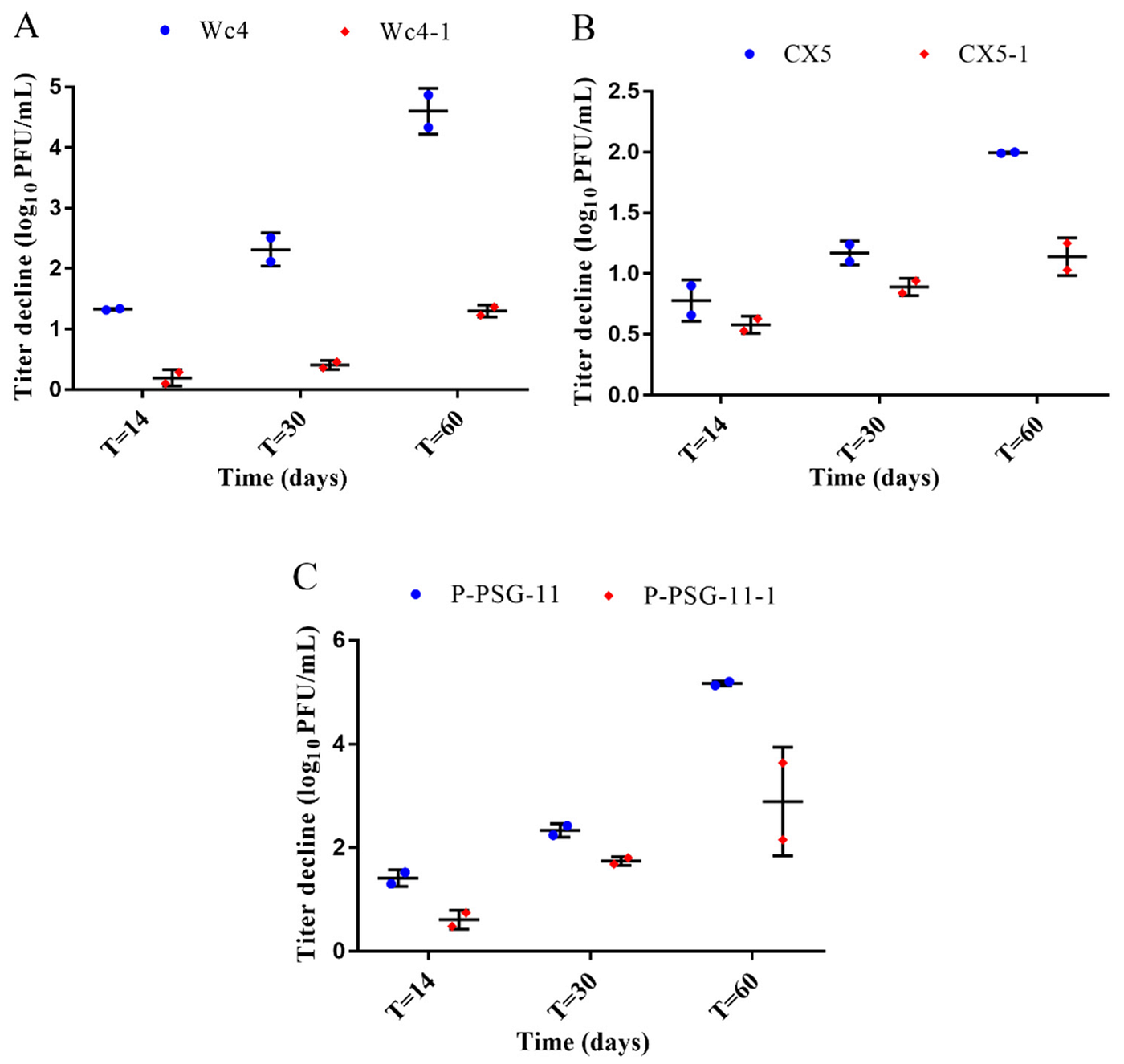
Results pulled from “Application of Adaptive Evolution to Improve the Stability of Bacteriophages during Storage” depicting the titer reduction of the ancestral phages (blue) and the heat-treated phages (red) after 60 of incubation at 37℃. Results are the mean values from two different experiments.
Mutations of these genomes only occurred in coding regions, and there were no insertions/deletions observed⁴. This means that the phages themselves evolved and made coding changes to their genomes as they were experiencing heat exposure, and there weren’t any new gene sequences that were deleted/inserted into their genome entirely. Wc4–1 had a mutation in a putative protein (function is unknown). Phage CX5–1 had a mutation in its tail tubular protein gp12⁴. This gene is involved in phage structure and infection. Phage P-PSG-11–1 had a mutation in its tail tubular protein gp11, also responsible for phage structure and infection. Overall, no major structural changes occurred. The cytosine nucleotide showed the most mappable mutations in the phage genomes⁴.
This paper shows that adaptive evolution and harnessing naturally occurring events like species-driven improvements after being exposed to alternative environments is also a technique for genetically modifying phages without the need for artificial modifications. This technique will be especially crucial for modifying phages’ stability levels and storage ranges.
Modular Receptor-Binding Proteins (RBPs) and Serotype Specificity
One of the restrictions in utilizing phage therapy is how niche phages are. This is also a benefit because it limits the amount of interference it will have on the body, but it also limits the variety of where we can use the phages. That’s why there has been research in using gene editing to modify the receptor-binding proteins phages have so they can have broader host ranges and bind to different strains of bacteria. In this experiment, scientists were looking at how they could modify the receptor-binding proteins of Klebsiella phages to adapt to multiple variations of the Klebsiella bacterial strain⁵. There are two main components to receptor-binding proteins (RBPs). There are N-terminal structural modules, which are responsible for attaching the RBP to the phage tail, as well as C-terminal specificity modules, which are responsible for recognizing and degrading specific capsular polysaccharides. In the experiment, in order to attempt to modify the RBPs, they constructed modular chimeras by swapping the N-terminal and C-terminal modules between phages, and they created synthetic RBPs comprised of multiple different components. This process mimicked natural horizontal gene transfer, which is when genetic material is transferred between different organisms to gain new abilities, which is an occurrence that might happen during phage evolution. The ability to assemble the RBPs was done with the VersaTile technique, which involved creating “tiles” from the coding sequences. Then, for phage genome assembly, multiple genome fragments were constructed with yeast cells using a technique called yeast gap repair, which is used to join multiple DNA fragments into a complete genome. Once the entire genome is constructed into one entire synthetic phage genome with the N-terminal and C-terminal modifications, that genome is extracted from the yeast and purified into DNA. Then, the DNA was either stored as a circular plasmid or as linear DNA. Whatever it may have been, that purified DNA was electroporated into an E. Coli strain where it was rebooted. Once inside the E. Coli strain, the synthetic genome began expressing its genes and the phage life cycle was activated, producing functional phages with the desired genome and modifications⁵.
Klebsiella phages KP32, KP34, and KP36 were used in this study to provide different C-terminal modules and N-terminal modules⁵. KP32 was made up of a first RBP (1A), which connects to the phage tail, a second RBP (2E), which interacts with the first RBP, and de-polymerase domains (proteins with the ability to break down the protective layers on the surface of bacteria) 1E and 2E which are specific to K3 and K21 Klebsiella bacterial strains (also referred to as serotypes). KP34 was made up of a single RBP (3E), which was indirectly attached to the phage via an intermediate anchor protein (3A), with specificity to the K63 serotype, and has a de-polymerase domain 3E. For phage KP36, it had a single RBP (4E) attached directly to the phage particle via an anchor domain (4A), specificity to the K63 serotype, and de-polymerase domain 4E. They utilized the components to create chimeras that derived from the anchor (1A, 3A, and 4A) and enzymatic (1E, 2E, 3E, and 4E) domains of these 3 phages to create 12 different chimera combinations. 4 other chimera combinations were created using an anchor domain from phage K11 combined with the enzymatic domains, making a total of 16 different chimera phages constructed⁵.
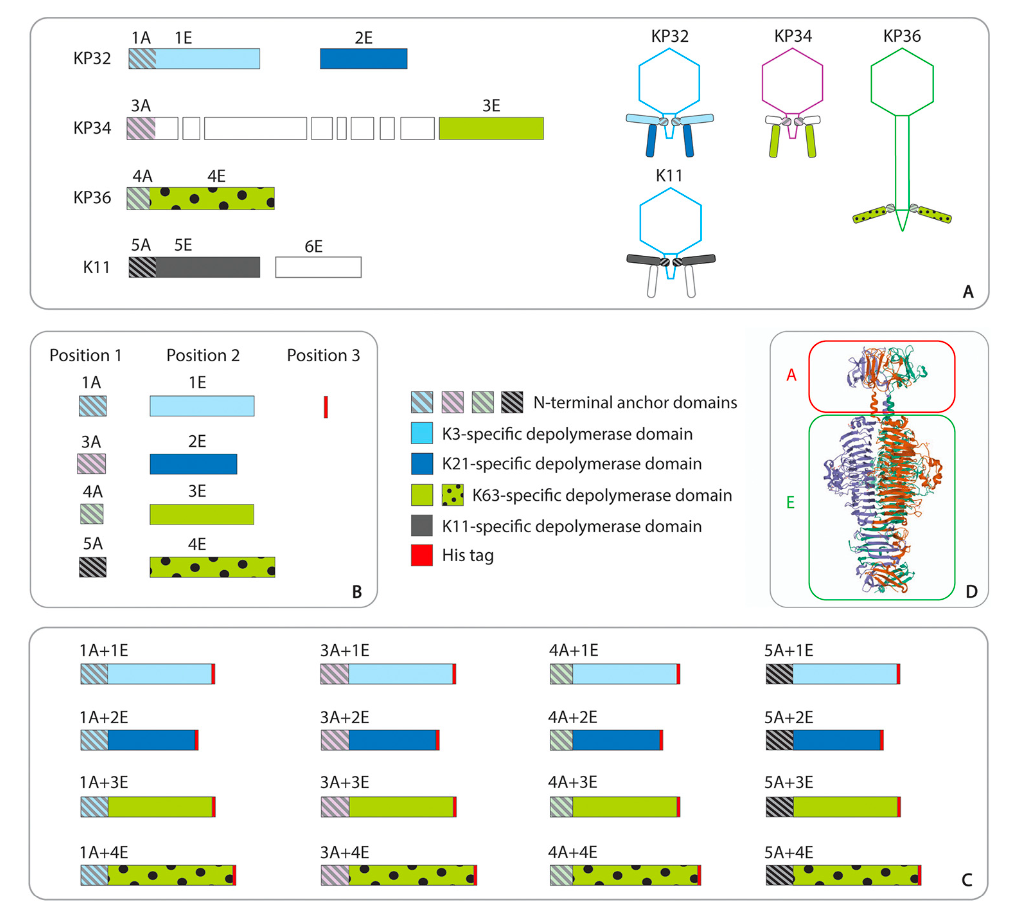
Diagram pulled from “Engineering the Modular Receptor-Binding Proteins of Klebsiella Phages Switches Their Capsule Serotype Specificity” depicting the modular architecture of the Klebsiella phage RBPs and their chimeras used in the experiment. Photo credit
The different combinations resulted in specificity shifts of the phages between the different serotypes K3, K21, and K63 based on the anchor and enzymatic domain the phage had⁵. For example, phage K115A1E (K11 anchor + KP32 enzymatic domain) switched specificity to K3 serotype, phage K115A2E (K11 anchor + KP32 2E enzymatic domain) switched to K21 serotype, and phage K115A3E (K11 anchor + KP34 3E enzymatic domain) shifted specificity to K63 serotype.⁵
This shows the capabilities for swapping the host range of phages for more versatile use and overcoming the limits in niche phage hosts, also providing the possibility to increase host ranges in phages and allow them to have broader use in phage therapy.
Discussion
While the field combining phage therapy and genetic engineering is evolving at a rapid pace, there are still obstacles that need to be considered when deciding if phage therapy should be FDA-approved and used clinically. Phages will not be able to be consistently produced as a medical treatment without perfecting gene editing techniques. The production of phages will need to be precise, consistent, and scalable. If we are going to rely on phages over antibiotics completely or as co-treatments, we will have to be able to harness and produce both a large amount and a large variety of phages. This will ensure that the phages can be utilized as an effective treatment at any time without anybody having to wait too long for treatment access, as well as have phage therapy be effective for any strain of bacteria, because phages can only infect a niche amount of bacteria based on their compatible RBPs.
Side-by-side recap of gene editing techniques for genetically engineering phages. Top left: homologous recombination and plasmid-based recombination. Requires there to be two donor DNA templates with homologous (relating) characteristics. in plasmid-based recombination, a donor plasmid is provided, which stores the necessary components for gene editing to occur. Gene editing happens naturally, therefore it is not guaranteed that the edits will be accurately made. Top right: PHEIGES method. PHEIGES method takes already genetically engineered phages and multiplies their quantities for easier scaling of phage therapy treatments. The procedure takes about ~3 days (actual amplification and assembly takes ~1 day). Bottom left: CRISPR-Cas gene editing method. CRISPR-Cas method is more precise and less dependent on multiple resources compared to other treatments. Has a higher success rate, cost-effective, efficient, and widely used compared to other methods. Is recommended for use for genetically engineering phages. Bottom center: CRISPR-Cas9 gene editing method for use in phages. The same method happens within a bacterial host for easier access to DNA for editing as well as reconstruction of newly edited phages afterwards. The Cas9 method is the most commonly used out of all the Cas systems available. Bottom right: CRISPR-Cas12a gene editing method. Has been recently researched to be more effective compared to the more commonly used Cas9 method. has the same process as the CRISPR-Cas9 procedure. Not listed in the diagram: BRED gene editing method. Enhanced homologous recombination occurs between two DNA sequences with similar components. The enhancement occurs by either utilizing already phage-encoded recombination systems or, if not present in phages, a recombination protein system that is introduced via plasmid into the bacterial host where the edits are taking place. Has limited use in Gram-positive bacteria. The method also demonstrates low transformation efficiencies between the DNA strands for editing.
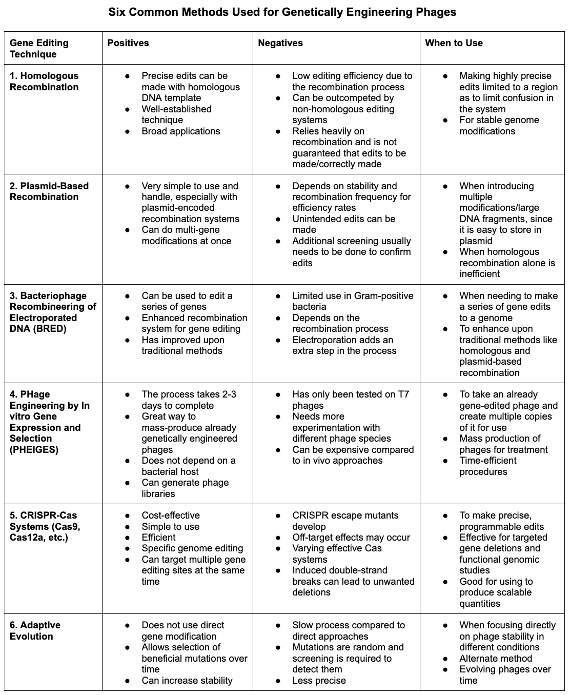
The table depicts the different gene editing techniques discussed, showing their positives, negatives, and when to use each technique.
One of the problems that experts are facing in gene editing phages is the occurrence of escape mutants. Escape mutants most commonly appear when using CRISPR-Cas gene editing systems, which has become a popular gene editing technique. Escape mutants are when phages mutate their own DNA to evade CRISPR targeting¹. Because the phage mutated its own DNA, CRISPR can no longer bind and cut the region in the phage genome that was originally being targeted. That phage can then escape the gene editing occurrence. If this keeps happening, there can be more and more stalls when creating a stock of phages for clinical use⁹. One solution to this is to design multiple gRNAs that target different regions of the same gene. This approach can ensure that if a mutation occurs by the phage in one target site, the other target sites in the gene can still be recognized by the other gRNAs and be cleaved. This still works because it is only required that one site in a gene be targeted and cleaved for the rest of the gene editing process to work. So even if there is a mutation that occurs in one site, the other sites can still be cleaved by the other gRNAs.
The methods for phage engineering discussed in this paper are all still being researched and further improved upon, with the goal of enhancing the ability to engineer phages at scale for mass production. Gene editing techniques like CRISPR-Cas systems are the modern version of other past utilized gene editing methods and are getting the most interest right now in this field. CRISPR-Cas systems are the techniques that are currently the quickest and most scalable for use. There has also been more research in using different cas-systems that differ from the more commonly used Cas-system, Cas9. Other Cas systems include Cas10, Cas12, Cas13, and others, which have been researched specifically for phage therapy application to provide techniques that will be able to mass produce phage therapy treatment¹. While there is research being done for CRISPR-Cas techniques, there is still constant improvement upon older methods such as homologous recombination, plasmid-based recombination, BRED, and others that weren’t explicitly mentioned in this paper, in order to keep finding new breakthroughs that will help combat the obstacles in implementing phage therapy, which gene editing can solve.
As well as utilizing a multitude of gene editing methods, which is necessary in order to make phage therapy a suitable treatment option, there has been exploration into other tools such as PHEIGES, discussed in this paper, to help with the mass production and scale of genetically engineered phages. This method will be one that helps take pre-engineered phages and produce them at a fast rate in large quantities to ensure there is constant phage stock that can be accessed whenever it is necessary and not jeopardize patient safety while they are waiting for those life-saving treatments. PHEIGES will become crucial alongside gene editing techniques like CRISPR to make phage therapy accessible to all and become a usable clinical treatment.
Looking into more unique tools such as adaptive evolution will also become an important technique in commercializing phage therapy for clinical applications. Adaptive evolution may become a common tool alongside CRISPR in improving phage stability both in treatment and during storage in order for them to stay functional under various stress conditions. Using this method and focusing on their stability will also be able to broaden phage therapy applications outside of the human body. There has been research on applying phage therapy for veterinary medicine (briefly covered in this paper), crops and agriculture, and even drug/gene carriers.
Not covered in this paper, there has been much research on using phages as delivery systems for other treatments like antibiotics, drugs, and even other genetic material, to be administered straight to a source. This broadens the use of phage therapy even more as it does not require the phage to specifically be compatible with the target host, as there is now the possibility that their RBPs can be modified to recognize a broader range of targets that stretch far beyond bacterial hosts, such as possibly cancerous cells or other conditions. Genetic engineering of the RBPs also allows for more personalization of phage therapy treatment, which is one of the advantages to using this treatment as it, in theory, does not interfere with the parts of the body but only targets the desired location that is causing a problem. This aligns with the future directions of healthcare and human longevity, proving that phage therapy is not an outdated method but rather one that is unexplored.
Conclusion
The advancements being made for combating antibiotic resistance by utilizing phage therapy are accelerating greatly. Combining the use of genetic engineering has played a huge role in phage therapy breakthroughs in the last few years, and it is the key to overcoming the current obstacles that implementing phage therapy faces and making this treatment one that can be accessible to all in a timely manner. Utilizing gene editing techniques like CRISPR-Cas systems as well as tools like PHEIGES for scalable phage therapy production, while consistently improving upon older methods like homologous recombination and others, is the future of phage therapy. Phage therapy in clinical applications is considered to be the world’s final solution to antibiotic resistance, and it must keep progressing with the use of gene editing to prevent the antibiotic resistance crisis from becoming a global problem that the whole world may face.
References
¹Chen, Y., Batra, H., Dong, J., Chen, C., Rao, V. B., & Tao, P. (2019). Genetic engineering of bacteriophages against infectious diseases. Frontiers in Microbiology, 10. https://doi.org/10.3389/fmicb.2019.00954
²Chen, Y., Yan, B., Chen, W., Zhang, X., Liu, Z., Zhang, Q., Li, L., Hu, M., Zhao, X., Xu, X., Lv, Q., Luo, Y., Cai, Y., & Liu, Y. (2024). Development of the CRISPR-Cas12a system for editing of Pseudomonas aeruginosa phages. iScience, 27(7), 110210. https://doi.org/10.1016/j.isci.2024.110210
³Cong, X., Zhao, S., Zhang, Q., Liu, S., Zhang, Y., & Yan, F. (2024). Isolation, Characterization, and Genome Engineering of a Lytic Pseudomonas aeruginosa Phage. Microorganisms, 12(11), 2346. https://doi.org/10.3390/microorganisms12112346
⁴Kering, K. K., Zhang, X., Nyaruaba, R., Yu, J., & Wei, H. (2020). Application of Adaptive Evolution to Improve the Stability of Bacteriophages during Storage. Viruses, 12(4), 423. https://doi.org/10.3390/v12040423
⁵Latka, A., Lemire, S., Grimon, D., Dams, D., Maciejewska, B., Lu, T., Drulis-Kawa, Z., & Briers, Y. (2021). Engineering the modular Receptor-Binding proteins of Klebsiella Phages switches their capsule serotype specificity. mBio, 12(3). https://doi.org/10.1128/mbio.00455-21
⁶Levrier, A., Karpathakis, I., Nash, B., Bowden, S. D., Lindner, A. B., & Noireaux, V. (2024). PHEIGES: all-cell-free phage synthesis and selection from engineered genomes. Nature Communications, 15(1). https://doi.org/10.1038/s41467-024-46585-1
⁷Nobrega, F. L., Costa, A. R., Santos, J. F., Siliakus, M. F., Van Lent, J. W. M., Kengen, S. W. M., Azeredo, J., & Kluskens, L. D. (2016). Genetically manipulated phages with improved pH resistance for oral administration in veterinary medicine. Scientific Reports, 6(1). https://doi.org/10.1038/srep39235
⁸World Health Organization: WHO. (2023, November 21). Antimicrobial resistance. Retrieved February 7, 2025, from https://www.who.int/news-room/fact-sheets/detail/antimicrobial-resistance
⁹Zhang, X., Zhang, C., Liang, C., Li, B., Meng, F., & Ai, Y. (2022). CRISPR–CAS9 based bacteriophage genome editing. Microbiology Spectrum, 10(4). https://doi.org/10.1128/spectrum.00820-22
메타데이터
- post_id
- 9c75a816720b
- slug
- methods-and-applications-for-genetically-engineering-phages-to-further-phage-therapy-progress-and-9c75a816720b
- url
- https://medium.com/@IsavellaT/methods-and-applications-for-genetically-engineering-phages-to-further-phage-therapy-progress-and-9c75a816720b
- canonical_url
- https://medium.com/@IsavellaT/methods-and-applications-for-genetically-engineering-phages-to-further-phage-therapy-progress-and-9c75a816720b
- author_url
- https://medium.com/@IsavellaT
- status
- ok
- fetched_at
- 2026-07-21 01:33:17