Why the Same CT Scan Can Look Completely Different
Understanding Window Width and Window Level in medical imaging.
Why the Same CT Scan Can Look Completely Different
Understanding Window Width and Window Level in medical imaging.
Photo by Umanoide on Unsplash
Imagine two radiologists looking at the exact same CT scan.
One is examining the lungs.
The other is looking for a small brain hemorrhage.
Surprisingly, they may not be looking at the same image.
Well… they are looking at the same CT scan, but not the same appearance of that scan.
How is that possible?
The answer lies in two simple concepts:
Window Width and Window Level.
Wait… Isn’t There Only One Image?
When a CT scanner acquires an image, it doesn’t produce a picture that’s ready to display.
Instead, it records thousands of pixel values representing tissue density.
Those values contain far more information than your monitor can display at once.
To make the image useful, we need to decide which range of values should be visible.
That’s exactly what windowing does.
Think About a Camera
Imagine taking a photo outside on a sunny day.
If you expose the image for the bright sky, the shadows become almost black.
If you expose it for the shadows, the sky becomes too bright.
The camera isn’t changing the scene.
It’s changing how that scene is displayed.
Windowing works in a similar way.
The CT data stays exactly the same.
Only its appearance changes.
What Is Window Level?
Window Level (WL) determines the center of the brightness range being displayed.
Think of it as choosing which tissue you want to focus on.
Different organs have different density ranges.
By changing the Window Level, you shift your attention toward a particular type of tissue.
What Is Window Width?
Window Width (WW) determines how wide that range is.
A narrow window increases contrast, making subtle differences easier to see.
A wider window displays a broader range of densities but with less contrast.
Neither setting is “better.”
They simply highlight different details.
One CT Scan, Multiple Views
A chest CT can be displayed using different window settings depending on what you’re looking for.
For example:
- Lung window highlights the lungs and airways.
- Soft tissue window emphasizes organs and muscles.
- Bone window makes bones much easier to evaluate.
The image data hasn’t changed.
Only the way it’s displayed has.
That’s why radiologists can switch between different windows with a single click.
Does Windowing Modify the DICOM File?
No.
This is an important distinction.
Changing the window does not modify the image stored in the DICOM file.
The original pixel values remain exactly the same.
Windowing is simply a different way of displaying those values.
Think of adjusting the brightness on your monitor.
The file doesn’t change — only what you see on the screen.
Where Are These Values Stored?
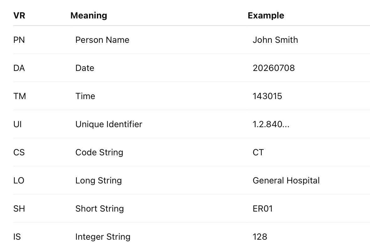
Many DICOM files include recommended window settings in their metadata.
For example:
- Window Center
- Window Width
These values help viewers display the image with sensible defaults.
Users can usually adjust them interactively if needed.
Viewing Window Settings with Python
Using pydicom, you can inspect these values.
import pydicomds = pydicom.dcmread("CT_Chest.dcm")print(ds.WindowCenter)
print(ds.WindowWidth)These values are often used by DICOM viewers when the image is first opened.
Why Developers Should Care
If you’re building a medical imaging application, simply displaying the raw pixel values isn’t enough.
Without proper windowing:
- Bones may appear too bright.
- Soft tissues may lose important detail.
- Lung structures may become difficult to interpret.
Supporting Window Width and Window Level is essential for creating a viewer that clinicians can actually use.
Key Takeaways
- CT images contain far more information than a monitor can display at once.
- Window Level determines the center of the displayed range.
- Window Width determines how much of that range is visible.
- Different window settings emphasize different tissues.
- Changing the window affects only the display, not the underlying DICOM data.
Final Thoughts
One of the things that makes CT imaging so powerful is that a single scan can reveal different kinds of information without acquiring new images.
By simply adjusting Window Width and Window Level, radiologists can examine the lungs, bones, or soft tissues — all from the same dataset.
It’s a great reminder that in medical imaging, what you see often depends on how you choose to look at it.
Before you go
- Please take a moment to like the post and follow the writer!
- Did you know that over 400,000 developers share what they’re building, learning, and discovering across our platforms every month? Learn how you can contribute here
메타데이터
- post_id
- a02a26c415ce
- slug
- why-the-same-ct-scan-can-look-completely-different-a02a26c415ce
- url
- https://blog.stackademic.com/why-the-same-ct-scan-can-look-completely-different-a02a26c415ce
- canonical_url
- https://blog.stackademic.com/why-the-same-ct-scan-can-look-completely-different-a02a26c415ce
- author_url
- https://medium.com/@koka-tic
- status
- ok
- fetched_at
- 2026-08-07 13:09:39