Top Mistakes to Avoid in Veterinary Dental Radiography Positioning
Veterinary dental radiography is one of the most valuable diagnostic tools in veterinary dentistry, yet many diagnostic errors occur before…
Top Mistakes to Avoid in Veterinary Dental Radiography Positioning
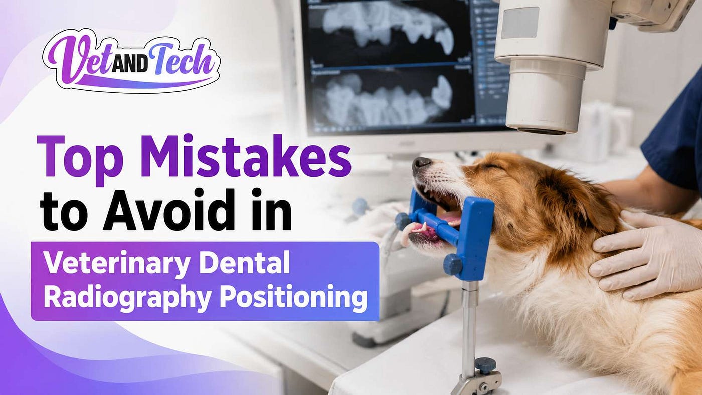
Veterinary dental radiography is one of the most valuable diagnostic tools in veterinary dentistry, yet many diagnostic errors occur before a veterinarian ever reviews the image. The problem is often not the equipment. It is positioning.
Studies have shown that a significant percentage of dental pathology in dogs and cats exists below the gumline and cannot be identified through visual examination alone. When radiographs are improperly positioned, clinicians risk missing tooth root abscesses, periodontal disease, tooth resorption, retained roots, and other painful conditions.
The good news is that most positioning errors are preventable. Understanding the common mistakes and how to correct them can dramatically improve image quality, diagnostic confidence, and patient outcomes.
Whether you are a veterinarian, veterinary technician, or dental assistant, mastering Veterinary Dental Radiography positioning is one of the fastest ways to improve your dental workflow and elevate patient care.
Why Is Proper Veterinary Dental Radiography Positioning So Important?
Dental radiographs are considered the standard of care in modern veterinary dentistry because many oral diseases occur beneath the gingiva where they cannot be seen during an oral examination.
According to the American Veterinary Dental College (AVDC), dental radiographs are essential for diagnosing and treating oral disease in companion animals. Missing even a small area due to positioning errors can lead to incomplete diagnoses and delayed treatment.
Proper positioning helps clinicians:
- Capture the entire tooth and surrounding structures
- Evaluate periodontal bone loss accurately
- Detect root fractures and retained roots
- Identify tooth resorption lesions
- Improve treatment planning
- Reduce retakes and anesthesia time
In busy veterinary practices, reducing repeat exposures also improves efficiency and minimizes patient risk.
What Are the Most Common Veterinary Dental Radiography Positioning Mistakes?
1. Incorrect Sensor Placement
Poor sensor placement is one of the most frequent causes of diagnostic image failure.
When the sensor is positioned too far rostrally, caudally, dorsally, or ventrally, important anatomy may be excluded from the image.
Common signs of poor sensor placement include:
- Missing root apices
- Cropped crowns
- Incomplete periodontal structures
- Distorted anatomy
A useful rule is to position the sensor so that the entire tooth and surrounding alveolar bone are captured within the image field.
2. Improper Beam Angle Selection
Beam angulation directly affects image accuracy.
In veterinary dentistry, the bisecting angle technique is commonly used because intraoral anatomy often prevents parallel positioning.
When the x-ray beam is incorrectly angled:
- Teeth appear elongated
- Teeth appear shortened
- Root structures become distorted
- Pathology can be hidden
Dr. Brett Beckman, DVM, FAVD, DAVDC, a board-certified veterinary dentist, frequently emphasizes that understanding bisecting angle geometry is one of the most important skills for producing diagnostic dental radiographs.
3. Failure to Visualize Root Apices
One of the biggest diagnostic mistakes is obtaining an image that does not include the root apex.
Many significant dental conditions occur at or near the apex, including:
- Periapical abscesses
- Endodontic disease
- Root fractures
- Inflammatory lesions
Without complete visualization, clinicians may overlook clinically significant pathology.
Before exposing the image, always verify that the sensor extends beyond the anticipated root length.
Why Are Maxillary Teeth Often More Difficult to Position?
Maxillary teeth present unique anatomical challenges.
The skull shape of dogs and cats can make proper sensor placement difficult, particularly for:
- Maxillary fourth premolars
- Maxillary molars
- Canine teeth
- Incisors
The roots of these teeth often extend in multiple directions, requiring precise positioning and beam alignment.
For this reason, many veterinary teams spend additional training time mastering maxillary radiographic techniques.
One of the best ways to improve these skills is through continuing education focused specifically on dental radiography positioning.
Veterinary professionals looking for hands-on instruction can benefit from the specialized webinar offered through Vet and Tech covering dental radiography positioning techniques taught by experienced veterinary dental educators.
Are You Making Positioning Errors Without Realizing It?
Many positioning mistakes become habits over time.
Ask yourself these questions:
- Do I routinely need multiple retakes?
- Are root apices frequently missing?
- Do images appear stretched or compressed?
- Are crowns overlapping adjacent teeth?
- Do I struggle with maxillary premolars and molars?
If the answer is yes to any of these questions, positioning technique may be limiting image quality.
Continuous training can help identify blind spots and improve consistency.
How Can Veterinary Teams Improve Dental Radiography Positioning Skills?
The most effective approach combines practice, education, and structured feedback.
Best Practices for Better Positioning
- Follow a consistent imaging protocol.
- Learn bisecting angle principles thoroughly.
- Review radiographs immediately after exposure.
- Analyze positioning errors before retaking images.
- Participate in continuing education programs.
- Practice full-mouth radiographic series regularly.
- Learn species-specific positioning variations.
Research published in veterinary dental education literature consistently demonstrates that structured training significantly improves radiographic quality and diagnostic accuracy.
What Can Veterinary Technicians Learn from Advanced Dental Imaging Training?
Veterinary technicians play a critical role in obtaining diagnostic-quality radiographs.
Advanced training helps technicians:
- Improve positioning accuracy
- Reduce anesthesia time
- Increase diagnostic yield
- Improve workflow efficiency
- Support better patient outcomes
Education is especially important as veterinary dentistry continues to evolve and expectations for diagnostic standards increase.
For professionals seeking practical instruction, the webinar Dental Radiography Positioning with Perrone provides focused guidance on positioning techniques, image interpretation considerations, and common troubleshooting strategies encountered in clinical practice.
The webinar is available through Vet and Tech’s Dental Radiography Positioning with Perrone Program and is designed for veterinarians, technicians, students, and other veterinary professionals looking to strengthen their dental imaging skills.
Where Can Veterinary Professionals Earn Free CE Credits While Improving Dental Skills?
Continuing education remains one of the most effective ways to stay current with evolving standards in veterinary medicine.
In addition to dental radiography education, Vet and Tech provides veterinary learning resources, educational content, and opportunities to participate in free continuing education programs.
Professionals interested in expanding their knowledge can explore available educational resources and register for upcoming learning opportunities, including various **Free CE upcoming veterinary webinars** that cover dentistry, internal medicine, wellness, diagnostics, leadership, and emerging veterinary topics.
These programs help veterinary teams maintain clinical excellence while earning valuable continuing education credits.
Frequently Asked Questions About Veterinary Dental Radiography
What is the most common mistake in veterinary dental radiography?
Incorrect sensor positioning is the most common error. It often results in missing root structures and nondiagnostic images.
Why are dental radiographs important in veterinary medicine?
Most dental disease occurs below the gumline. Radiographs help identify pathology that cannot be detected during visual examinations.
What causes elongation on a dental radiograph?
Elongation occurs when the x-ray beam angle is too shallow relative to the bisecting angle.
What causes foreshortening on a dental radiograph?
Foreshortening occurs when the beam angle is too steep, causing the tooth image to appear compressed.
How can veterinary technicians improve radiography positioning?
Regular practice, structured continuing education, image review, and advanced dental radiography training are among the most effective methods.
Final Expert Insight
The difference between an average dental radiograph and a diagnostic-quality image often comes down to millimeters of positioning and a few degrees of beam angulation. Every missed apex, distorted root, or overlapping crown creates an opportunity for disease to go unnoticed. As veterinary dentistry continues to advance, positioning skills are becoming just as important as interpretation skills. The teams that invest in education, refine their techniques, and consistently review their images are the ones most likely to identify disease earlier, improve patient comfort, and deliver a higher standard of care.
Sources
- Vet and Tech — Dental Radiography Positioning with Perrone Webinar: https://www.vetandtech.com/webinars/dental-radiography-positioning-with-perrone
- American Veterinary Dental College (AVDC) — Dental Radiography Guidelines.
- American Animal Hospital Association (AAHA) Dental Care Guidelines for Dogs and Cats.
- Niemiec BA. Veterinary Dentistry Clinical Practice and Procedures.
- Beckman BW, DVM, FAVD, DAVDC — Veterinary Dental Radiography Education Resources.
- World Small Animal Veterinary Association (WSAVA) Dental Guidelines.
메타데이터
- post_id
- a63769ac5d2a
- slug
- top-mistakes-to-avoid-in-veterinary-dental-radiography-positioning-a63769ac5d2a
- url
- https://medium.com/@Veterinary_resources/top-mistakes-to-avoid-in-veterinary-dental-radiography-positioning-a63769ac5d2a
- canonical_url
- https://medium.com/@Veterinary_resources/top-mistakes-to-avoid-in-veterinary-dental-radiography-positioning-a63769ac5d2a
- author_url
- https://medium.com/@Veterinary_resources
- status
- ok
- fetched_at
- 2026-07-22 05:21:28