Introduction to CT DICOM Images (Part 1)
Computed Tomography (CT) scans are widely used in medical diagnosis and research. They help doctors see what is happening inside the human…
Introduction to CT DICOM Images (Part 1)
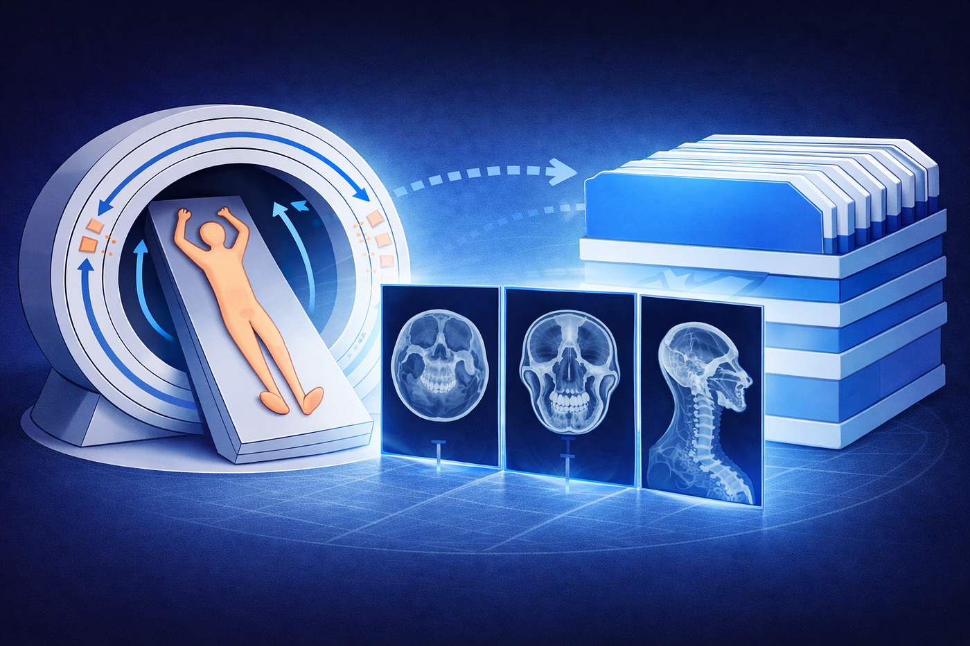
AI-generated image
Computed Tomography (CT) scans are widely used in medical diagnosis and research. They help doctors see what is happening inside the human body, layer by layer.
At first glance, working with CT data sounds simple until you actually open a CT dataset.
When I first opened a CT dataset, I expected to see a few image files. Instead, I found hundreds of files, all with the same unfamiliar format: DICOM.
This is the first surprise for anyone new to medical imaging.
CT data is not stored like normal images such as JPEG or PNG. Instead, a CT scan is stored as a series of DICOM files, where each file represents a small slice of the body.
To understand CT data properly, we must first understand what DICOM is.
What is DICOM?
DICOM stands for Digital Imaging and Communications in Medicine.
It is the standard format used in medical imaging to store, share, and manage medical images across hospitals, scanners, and software systems.
A DICOM file is not just an image. It is more like a package that contains both the image and important information about how that image was created.
What Does a DICOM File Contain?
A DICOM file contains two main parts, metadata and image data. You can think of it like a labeled photo album page one part explains the picture, and the other part is the picture.
Metadata (The “Labels”) Metadata is the information stored inside the file that describes the image It works like an ID tag attached to the scan. This information is stored in a standardized section called the DICOM header, which appears at the beginning of the file.
The metadata answers the important questions:
- Who was scanned (patient details)
- What was scanned (CT, MRI, scan settings)
- Where the image belongs in the body (slice position)
- How the image was captured (pixel spacing, slice thickness, scanner details)
Image Data (The “Picture”)
This is the actual image stored inside a DICOM file. It represents what the medical scanner captured from inside the body.
The image is made up of pixels. Each pixel does not store a color like a normal photo. Instead, it stores a numeric value that describes how bright or dark that small area should appear.
Where is DICOM Used?
DICOM is widely used in many medical imaging systems,
- CT (Computed Tomography)
- MRI
- Ultrasound
- X-ray
DICOM is a global standard that allows images from different machines and hospitals to work together seamlessly.
Now that we understand what DICOM is, let’s move back to CT imaging.
CT (Computed Tomography) is widely used in medical diagnosis because it allows doctors to see inside the body, layer by layer.
But how does CT actually work?
CT Working
AI-generated image
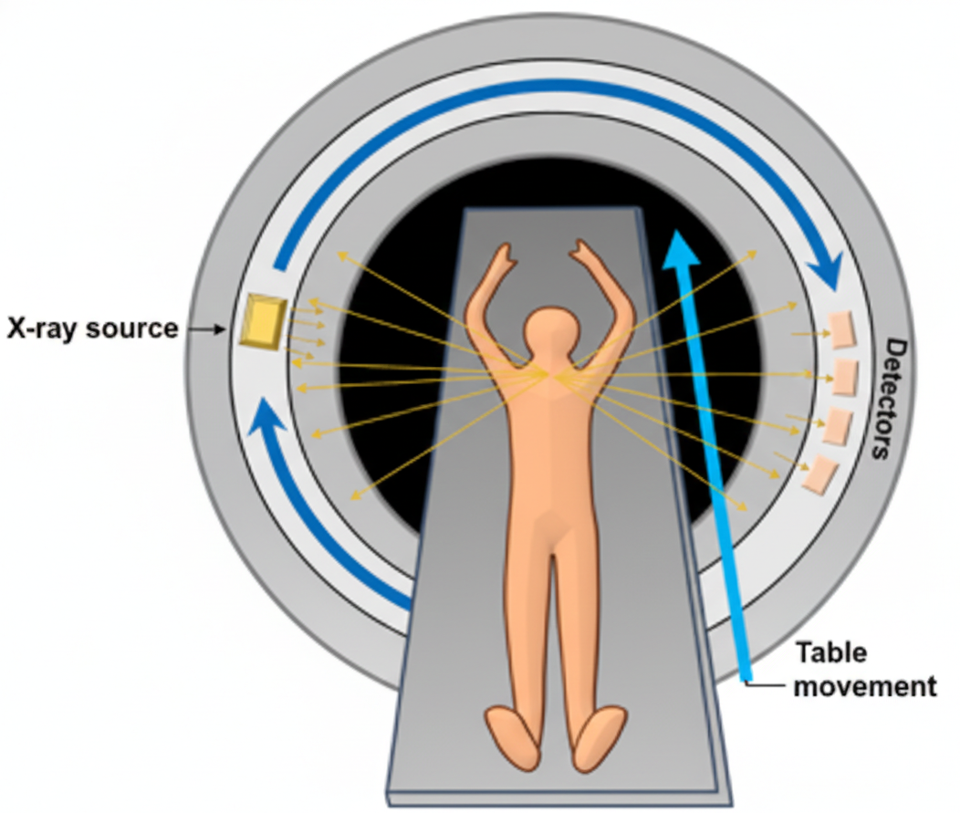
CT imaging works using X-rays to create detailed images of the human body. During the scan, the X-ray source rotates around the patient and sends X-rays through the body from multiple angles. Detectors placed on the opposite side collect the X-rays that pass through the body. Dense tissues such as bones absorb more X-rays, so fewer X-rays reach the detector, while air-filled regions allow more X-rays to pass through. These differences in X-ray attenuation are processed by a computer to generate cross-sectional images of the body.
Why Do CT Scans Have Hundreds of DICOM Files?
When you open a CT study in any DICOM viewer, you usually see three views.
- Axial
- Coronal
- Sagittal
When I first studied CT imaging, I wondered
How can one CT scan have three different views?
The answer is simple.
A CT scan is not a single image.
It is a collection of many images stacked together.
Stack of CT Slices
Think of a loaf of bread 🍞. Each slice of bread represents one image.
When you stack all the slices together, you get the complete loaf.
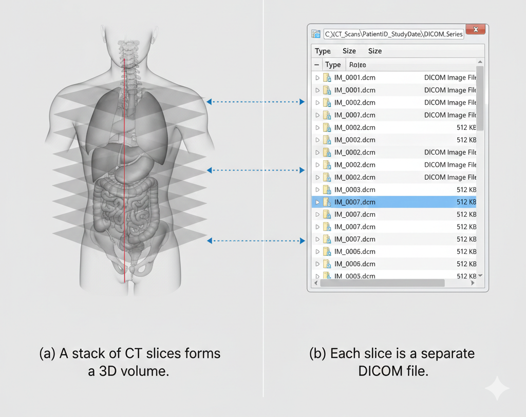
A CT scan works in a very similar way. Each DICOM file represents a single slice of the body. When all these slices are stacked together, they form a complete 3D volume of the body.
AI-generated image
How Do the Three Views Form?
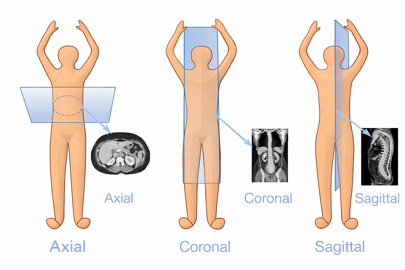
After all CT slices are stacked together, they form a 3D volume of the body. From this single volume, we can view the anatomy from different directions. The axial view shows the body from top to bottom, the coronal view from front to back, and the sagittal view from left to right.
- Axial view → looking from top to bottom
- Coronal view → looking from front to back
- Sagittal view → looking from left to right
AI-generated image
These are not separate scans. They are just different ways of looking at the same stacked data. This is why CT can show the body in three views using the same dataset.
In the next part, we will explore Hounsfield Units (HU), which describe tissue density in CT images. Understanding these values will help us see how bones can be identified and extracted from CT data.
메타데이터
- post_id
- b2bec89a3ecb
- slug
- introduction-to-ct-dicom-images-part-1-b2bec89a3ecb
- url
- https://medium.com/@ganeshan187/introduction-to-ct-dicom-images-part-1-b2bec89a3ecb
- canonical_url
- https://medium.com/@ganeshan187/introduction-to-ct-dicom-images-part-1-b2bec89a3ecb
- author_url
- https://medium.com/@ganeshan187
- status
- ok
- fetched_at
- 2026-06-23 06:34:20