The Hidden Universe: Learning to See with an Electron Microscope in Midjourney
“The most extraordinary landscapes are often the ones hidden beneath our fingertips.”
The Hidden Universe: Learning to See with an Electron Microscope in Midjourney
“The most extraordinary landscapes are often the ones hidden beneath our fingertips.”
The Breath of the Image
There is a strange moment that happens the first time you look through an electron microscope.
A grain of pollen suddenly resembles an alien fortress.
The surface of a butterfly wing becomes an endless mountain range.
A single strand of hair transforms into an architectural monument carved by time itself.
Nothing has changed.
Only your way of seeing.
This is perhaps one of the most fascinating aspects of MidJourney. It doesn’t simply imitate artistic styles. It can also imitate the visual language of scientific instruments, allowing us to explore worlds our eyes were never designed to perceive.
And among these instruments, few are as visually captivating as the electron microscope.
The Big Idea
When people write prompts, they usually describe what they want to generate, but experienced Midjourney users know that another question is often far more important:
How is this world being observed?
Changing the observer changes the entire image.
The camera is no longer just a camera, it becomes a scientific instrument, as an astronomical telescope, an X-ray machine or an electron microscope.
This subtle change completely transforms the visual grammar of the generated image.
What Is an Electron Microscope?
Unlike an optical microscope, which uses visible light, an electron microscope uses beams of electrons to reveal structures thousands, even millions of times smaller than what the human eye can perceive.
Instead of photographing reality, it reveals another one.
Tiny becomes monumental.
Texture becomes landscape.
Imperfections become architecture.
Objects stop looking familiar and begin looking almost extraterrestrial.
This is exactly why the style works so well inside Midjourney.
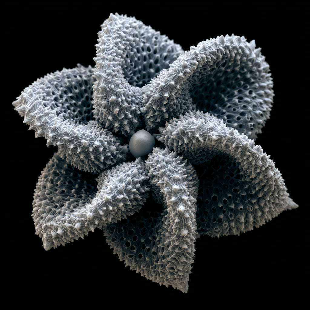
Mushroom spore captured with a scanning electron microscope, intricate sculpted shell covered in microscopic ridges and pores, surreal biological architecture, laboratory specimen, monochrome scientific imaging — ar 1:1 — raw — v 8.1
The Visual Translation
When Midjourney reads the words electron microscope, it doesn’t simply apply a grayscale filter.
It translates an entire scientific language.
That language includes:
extreme magnification
microscopic scale
dramatic surface texture
intricate geometric repetition
ultra-shallow spatial information
scientific lighting
laboratory precision
strong local contrast
biological complexity
material realism
You’re no longer asking MidJourney to create an object.
You’re asking it to reveal its invisible universe.
The Visual Alphabet
Every visual language is built from recurring elements.
The language of electron microscopy is no exception.
Magnification
Everything feels impossibly close.
Tiny details become entire landscapes.

petal from a crystalline fantasy flower viewed under a false-color scanning electron microscope, extreme magnification revealing vast mineral dunes, towering crystal formations and intricate cellular canyons stretching toward the horizon, violet, cyan, magenta and gold, scientific imaging — ar 16:9 — raw — v 8.1
Surface Texture
Smooth objects almost disappear.
Interesting objects become incredibly rich.
Wrinkles.
Fibers.
Pores.
Scales.
Crystals.
Nanostructures.

Specimen FY-034, enchanted fairy silk cocoon observed with a false-color scanning electron microscope, impossibly fine intertwined fibers woven into a dense three-dimensional network, delicate nanofilaments sparkling with turquoise, lavender, pearl white and gold, ultra-high magnification, breathtaking microscopic texture, laboratory realism — ar 4:5 — raw — v 8.1
Contrast
Electron microscopy is built around contrast.
Bright ridges.
Dark cavities.
Sharp transitions.

living crystal coral viewed through a false-color scanning electron microscope (SEM), enormous razor-like silica ridges emerging from labyrinthine shadow-filled cavities, microscopic mineral pores, brilliant cyan, emerald, magenta and gold, extreme local contrast, breathtaking nanostructure, publication-quality scientific micrograph — ar 4:5 — raw — v 8.1
Tiny shadows become dramatic.
Geometry
Nature suddenly looks engineered.
Honeycombs.
Hexagons.
Fractals.
Micro-architecture.

compound eye of a honeybee captured with a scanning electron microscope, thousands of perfectly arranged ommatidia forming a geometric dome, microscopic hairs between facets, extraordinary biological engineering, scientific imaging, high contrast, monochrome electron micrograph — ar 4:5 — raw — v 8.1
Repeating patterns appear everywhere.
Scale
Perhaps the most fascinating characteristic.
Without context, the viewer no longer knows whether they are looking at:
a mountain, or a grain of sand.

gravity moss examined through a false-color SEM, microscopic filament forests appearing as colossal valleys filled with towering vegetation, emerald, lime, turquoise and gold, no recognizable object size, extraordinary biological topography — ar 16:9 — raw — v 8.1
Scale completely disappears.
Why This Language Works So Well
Most prompts describe appearance.
Electron microscope prompts describe discovery.
They trigger curiosity.
The viewer instinctively wonders: “What am I looking at?”
That question creates engagement.
And curiosity is one of the strongest visual emotions.

quartz crystal fracture observed with a scanning electron microscope, microscopic crystalline terraces and angular formations, geological nano-landscape, laboratory SEM imaging, monochrome scientific realism, ultra sharp detail — ar 1:1 — raw — v 8.1

Specimen EL-006, leaf-like epidermal cell from an ancient forest elf examined through a false-color SEM, chlorophyll-inspired nanofibers forming elegant geometric mosaics, emerald, jade, turquoise and golden pollen deposits, scientific realism — ar 4:5 — raw — v 8.1
What This Language Changed in Me
One of the most surprising things about exploring scientific photography inside Midjourney is realizing that beauty doesn’t disappear as we zoom in.
It multiplies.
Every object hides another landscape.
Every surface contains another story.
The ordinary was never ordinary.
It was simply waiting to be observed differently.
Perhaps that’s what Midjourney teaches us best.
Creativity isn’t always inventing something new.
Sometimes it’s learning to look closer.
Libée Lune
메타데이터
- post_id
- c9646f71e39f
- slug
- the-hidden-universe-learning-to-see-with-an-electron-microscope-in-midjourney-c9646f71e39f
- url
- https://medium.com/@libelune/the-hidden-universe-learning-to-see-with-an-electron-microscope-in-midjourney-c9646f71e39f
- canonical_url
- https://medium.com/@libelune/the-hidden-universe-learning-to-see-with-an-electron-microscope-in-midjourney-c9646f71e39f
- author_url
- https://medium.com/@libelune
- status
- ok
- fetched_at
- 2026-06-22 17:31:34