Emerging Technologies in Optical Microscopy for Biomedical Applications
Deep learning in microscopy
Emerging Technologies in Optical Microscopy for Biomedical Applications

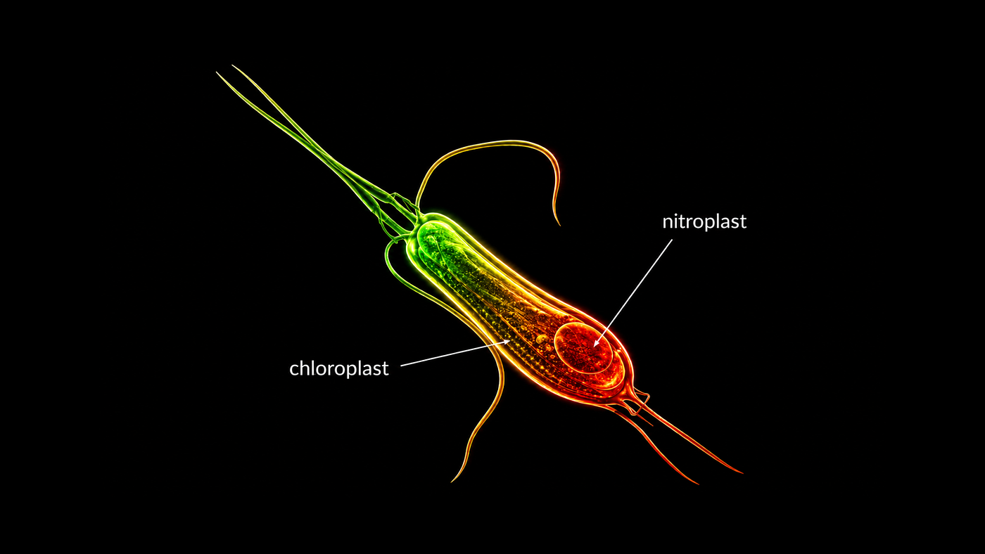
Digital painting illustration of optical microscopy
Optical microscopy has been one of the most basic technologies in biomedical research for centuries, enabling scientists to observe structures and biological processes that are too small to be seen by the human eye. Over the last few decades, microscopy has evolved from basic brightfield microscopes into sophisticated imaging platforms capable of providing detailed insights into cellular dynamics, molecular interactions, and tissue architecture. Nevertheless, conventional microscopy techniques have significant drawbacks, such as low contrast, diffraction-limited resolution, optical aberrations, phototoxicity, and limited imaging depth. These are even more important in live-cell imaging, where light exposure can harm the biological sample or change its natural behaviour. In recent years, quantitative phase microscopy, adaptive optical systems, and deep learning integration have emerged as new tools that are addressing many of these challenges and opening new avenues for biomedical imaging.
One of the most significant developments in modern microscopy is the introduction of quantitative phase microscopy (QPM), which allows label-free imaging of transparent biological samples. Traditional imaging methods like fluorescence microscopy require staining or fluorescent labelling to make the image more visible, but this can cause photobleaching and phototoxicity, which may affect the sample’s integrity. Unlike QPM, however, the intrinsic properties of cells can be analysed without the need for external labels, which is especially useful for observing living cells over long periods of time. Traditional label-free techniques such as phase contrast microscopy and differential interference contrast (DIC) microscopy improve image contrast, but they generally provide only qualitative information. QPM goes beyond these techniques by allowing the quantitative measurement of phase distributions, sample thickness, and refractive index variations.
Recent studies have shown that metasurfaces can be used to greatly improve QPM systems by replacing large optical components with planar structures. A multimodal optical differential system based on a dielectric metasurface has been developed to simultaneously support brightfield imaging, optical spatial differential imaging, differential interference contrast, and quantitative phase microscopy. The proposed system utilises the Pancharatnam–Berry phase to control light using sub-wavelength structures, which allows for the separation of the amplitude and phase information of complex optical fields. This innovation improves image contrast and enables single-shot phase acquisition without the need for complicated multi-directional alignment procedures that are commonly required in earlier phase reconstruction techniques. The technique was successfully demonstrated on paramecium cells, fishtail tissue and diatom cells.
At the same time, AI and deep learning are revolutionising the processing and analysis of microscopy data. Modern microscopes produce vast quantities of imaging data, and manual interpretation is becoming more and more challenging, time-consuming and inconsistent. Deep learning methods offer strong computational tools that can be used to automate image reconstruction, denoising, segmentation, classification and quantitative analysis. In optical microscopy, deep learning has emerged as a revolutionary tool to tackle issues like low signal-to-noise ratio, diffraction-limited resolution, and poor contrast. To enhance image quality and minimise reliance on human expertise, neural network architectures such as convolutional neural networks (CNNs), U-Nets, residual networks (ResNets), and generative adversarial networks (GANs) are now widely adopted.
The integration of deep learning in microscopy is especially useful for live-cell imaging and high-throughput experiments. In many biological studies, it is necessary to reduce the amount of light to preserve living samples, but this also reduces the quality of the signal and raises the noise level. Deep learning-based denoising methods can recover meaningful information from low-light images while minimising phototoxicity. Furthermore, automated segmentation and tracking systems allow for fast analysis of cellular structures and dynamic biological processes that would otherwise demand a lot of manual work. It has been demonstrated that deep learning can enhance imaging efficiency and make high-end imaging capabilities accessible to a broader audience, even in relatively simple imaging systems, through high-performance computational enhancement. There are, however, several issues that need to be addressed, such as the need for large annotated datasets, potential data bias, and the lack of interpretability of deep learning models.
The other trend in microscopy is the development of smart microscopy systems that actively adapt imaging conditions in real-time. In traditional microscopes, the acquisition parameters are set and chosen before the experiment. However, biological samples are dynamic, and imaging conditions need to be continually optimised to satisfy conflicting demands, including spatial resolution, acquisition speed, signal quality, and sample health. Smart microscopy is the combination of real-time analysis, feedback control and automated actuation. These systems modify imaging conditions during acquisition, based on information directly acquired from the specimen.
Smart microscopy tackles the “pyramid of frustration” in which researchers can enhance one imaging parameter at the expense of another. For instance, high spatial resolution often demands slower imaging times or less viable samples. Smart microscopy overcomes these limitations by tailoring imaging strategies to the experimental conditions. A microscope could be used at low temporal resolution for normal observation, and automatically switch to high-speed imaging only when rare biological events, such as mitosis, are detected. This adaptive strategy minimises the risk of phototoxicity and provides adequate detail of critical events. These systems have been rapidly developed, with the help of advances in automation, open-source microscope control software, and artificial intelligence, and are becoming more accessible to the scientific community.
The Multimodal Optical Scope with Adaptive Imaging Correction (MOSAIC) is one of the most advanced examples of multimodal microscopy, recently published in Nature Methods. MOSAIC combines multiple imaging modalities in a single reconfigurable platform, such as light-sheet microscopy, super-resolution imaging, multiphoton microscopy, and label-free imaging, all with adaptive optics correction. The system allows imaging at a variety of spatial and temporal scales, from single molecules to whole organisms, and provides high image quality even in complex multicellular environments. Importantly, MOSAIC permits switching between imaging modalities in the same experiment, allowing for comprehensive observation of biological processes with minimal disturbance to the sample.
The skills exhibited by MOSAIC are the future of biomedical imaging. The platform has been successfully applied to long-term volumetric imaging of live cultured cells for almost 24 hours, producing datasets of millions of image volumes with low phototoxicity. The system also allowed for the detailed observation of rare cellular events like tripolar mitosis, mitochondrial remodelling and dynamic membrane behaviour. MOSAIC is a perfect example of the growing integration, flexibility and intelligence of modern microscopy systems, combining adaptive optics, multimodal imaging and automated control.
To conclude, the combination of sophisticated optical engineering, adaptive microscopy, and artificial intelligence is transforming optical microscopy. Contrast enhancement and label-free quantitative analysis are being improved by quantitative phase microscopy and metasurface-assisted imaging, and deep learning is revolutionising image reconstruction and automated analysis. The creation of smart microscopy systems and multimodal platforms like MOSAIC is another example of how real-time adaptation and the combination of multiple imaging modalities can break through the long-standing trade-offs in biological imaging. These technologies will continue to develop and will offer increasingly accurate, non-invasive and information-rich visualisation tools that will greatly enhance biomedical research, diagnostics and our understanding of living systems.
메타데이터
- post_id
- ccddacbf8cfa
- slug
- emerging-technologies-in-optical-microscopy-for-biomedical-applications-ccddacbf8cfa
- url
- https://medium.com/@puntaindratomo/emerging-technologies-in-optical-microscopy-for-biomedical-applications-ccddacbf8cfa
- canonical_url
- https://medium.com/@puntaindratomo/emerging-technologies-in-optical-microscopy-for-biomedical-applications-ccddacbf8cfa
- author_url
- https://medium.com/@puntaindratomo
- status
- ok
- fetched_at
- 2026-06-09 14:34:10