Shape as Data: A Primer on Morphometrics and Landmarks
Every face, every skull, every fin — shapes carry biological stories. Morphometrics is the science of decoding those stories with numbers.
Shape as Data: A Primer on Morphometrics and Landmarks
Every face, every skull, every fin — shapes carry biological stories. Morphometrics is the science of decoding those stories with numbers.
When you glance at a friend across a crowded room, your brain instantly recognises them. It does not memorise coordinates or run calculations. It grasps the structure of their face as a whole. Morphometrics is our attempt to teach that same intuition to machines and mathematical frameworks.
The word itself is elegantly descriptive. Break it apart: morph, from the Greek for “shape,” and metrics, meaning numerical measurement. Together, morphometrics is the scientific study of biological shape. It is the art of turning curves, contours, and proportions into data that can be compared, analysed, and interpreted.
Morphometrics converts the geometry of living things into data, making the invisible variation between organisms visible and quantifiable.
The applications are broader than you might expect. Morphometrics sits at the intersection of biology, computer vision, and data science. It contributes to fields as varied as facial recognition, age estimation, expression analysis, orthodontics, evolutionary biology, bioinformatics, and species classification. And increasingly, it is powered not by human experts placing points by hand, but by neural networks doing it in milliseconds.
Two Generations of Morphometrics
The discipline splits into two broad methodological families: traditional morphometrics and geometric morphometrics.
Traditional morphometrics is the older approach. Researchers take physical measurements such as the width of a jaw, the height of a cranium, or the length of a fin, and use those distances and ratios as their data. It is intuitive and practical, but it has a fundamental weakness. Raw measurements do not fully capture shape. Two fish could share identical length measurements yet look completely different.
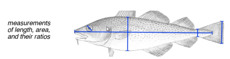
Figure 1. Traditional morphometrics in action: measurements of length, area, and their ratios taken on a cod. While straightforward, this approach captures isolated dimensions rather than overall form.
Geometric morphometrics emerged as a more powerful successor. Instead of isolated measurements, it captures the full geometry of an object through a set of carefully placed points called landmarks, or through traced outlines. These coordinates preserve the spatial relationships between all parts of a structure simultaneously, enabling richer and more nuanced comparisons.

Figure 2. Two approaches within geometric morphometrics: an outline traces the full boundary of the specimen (left), while landmarks mark a set of discrete, biologically meaningful points (right). Both preserve spatial relationships that traditional measurements discard.
Traditional morphometrics measures parts. Geometric morphometrics captures the whole.
Landmarks: The Vocabulary of Shape
In geometric morphometrics, a landmark is a specific point on an object that can be reliably matched to the equivalent point on every other object in your study. Think of it as a consistent reference system: the tip of the nose, the corner of the eye, the insertion point of a fin.
Not all landmarks are created equal. The field distinguishes between types based on how precisely and confidently a point can be identified across specimens. This reliability is captured by the concept of homology.
Homology, at its core, means “sameness.” A strongly homologous point is one you can identify on a hundred different specimens and say with confidence: yes, this is the same location in all of them. Weaker homology means greater uncertainty in that match and greater potential for error in your analysis.

Figure 3. Traditional landmarks applied to a fish (left) and anatomical landmarks plotted on a 3D facial model (right). The same principles of landmark placement apply across 2D and 3D data, though the tools and complexity differ substantially.
Four Types of Landmarks

Figure 4. Landmark types and their properties. Homology strength reflects how confidently a point represents the same location across all specimens in a study.
Anatomical landmarks are the gold standard. They are expert-defined points that correspond between organisms in a biologically meaningful way, such as the iris centre, the tip of the nose, or the corner of the mouth. Geometric landmarks are placed algorithmically at mathematically significant positions, like the point of greatest curvature along a bone edge. Constructed landmarks are derived from the positions of other landmarks; for example, the midpoint between two anatomical points. Finally, semi-landmarks are placed arbitrarily along a curve or surface and then optimised through a process called sliding semi-landmark analysis, which adjusts their positions via Procrustes analysis to best capture variation in outline shape.
This spectrum from anatomical to semi-landmark reflects a fundamental trade-off between biological precision and coverage. Anatomical landmarks are few but trustworthy. Semi-landmarks can densely sample an entire surface, but require optimisation to be analytically meaningful.
The Three Rules That Hold It All Together
For any landmark-based study to produce valid comparisons, three rules must be followed without exception:
Landmark consistency rules
- Every specimen must have the same number of landmarks.
- Landmarks must follow the same order. Landmark #7 must always represent the same anatomical location across every specimen.
- Landmarks must be placed on homologous points: locations that correspond across specimens based on shared morphology, function, or geometry.
Break any one of these rules and cross-specimen comparisons become meaningless. The entire statistical framework of geometric morphometrics rests on this consistency.
Scaling Up: From 2D to 3D
While classic morphometrics operated on photographs or physical specimens, modern tools now work directly on three-dimensional surface scans. A 3D facial model can carry hundreds of landmark points, and the same principles of homology and consistency apply at every one of them.

Figure 5. A 3D facial model covered in semi-landmarks (blue) and a smaller set of anatomical landmarks (red/orange), shown from frontal and lateral views. The dense blue cloud of semi-landmarks samples the overall surface geometry, while the anatomical anchors provide biological correspondence. This hybrid approach is increasingly common in modern facial morphometrics.
The contrast in the image above is striking: a handful of orange anatomical anchors surrounded by hundreds of blue semi-landmarks. The anatomical points provide biological grounding; the semi-landmarks fill in the geometric detail. Together, they give researchers a rich, high-resolution description of facial shape, the kind that supports applications in forensic identification, surgical planning, and facial recognition research.
Landmark Detection tools
Modern deep learning networks detect facial landmarks in milliseconds, consistently and at scale, without any human annotation.
MediaPipe Face Mesh predicts 468 landmarks across the full face surface. These are best understood as semi-landmarks: densely sampled, algorithmically placed points that capture overall surface geometry rather than discrete anatomical locations. MTCNN sits at the other end of the spectrum, returning just five points: the eye centres, nose tip, and mouth corners — each one a true anatomical landmark with strong biological homology. dlib’s 68-point predictor occupies the middle ground, combining anatomical anchors along the jawline, nose, and mouth with geometrically placed points along the brow and eye contours, giving it properties of both Type 1 and Type 2 landmarks.
References
Klingenberg, C. P. Introduction to Geometric Morphometrics. YouTube lecture series, PLnMoEqr0iq41Ec-bhkoZy-M93zVQvBYyJ. https://www.youtube.com/watch?v=3Soc75ox50A&list=PLnMoEqr0iq41Ec-bhkoZy-M93zVQvBYyJ&index=1 Zhang, K., Zhang, Z., Li, Z., and Qiao, Y. (2016). Joint Face Detection and Alignment Using Multitask Cascaded Convolutional Networks. IEEE Signal Processing Letters, 23(10), 1499–1503. Lugaresi, C. et al. (2019). MediaPipe: A Framework for Building Perception Pipelines. arXiv:1906.08172. King, D. E. (2009). Dlib-ml: A Machine Learning Toolkit. Journal of Machine Learning Research, 10, 1755–1758.
메타데이터
- post_id
- dd2efaf466a2
- slug
- shape-as-data-a-primer-on-morphometrics-and-facial-landmarks-dd2efaf466a2
- url
- https://medium.com/@sawaiz.naseem/shape-as-data-a-primer-on-morphometrics-and-facial-landmarks-dd2efaf466a2
- canonical_url
- https://medium.com/@sawaiz.naseem/shape-as-data-a-primer-on-morphometrics-and-facial-landmarks-dd2efaf466a2
- author_url
- https://medium.com/@sawaiz.naseem
- status
- ok
- fetched_at
- 2026-06-09 15:37:30