Why Some Bacteria Turn Purple and Others Turn Pink: The Science of Gram Staining
A step-by-step look at one of microbiology’s most essential lab techniques — and what it actually reveals about bacterial cell walls.
Why Some Bacteria Turn Purple and Others Turn Pink: The Science of Gram Staining
A step-by-step look at one of microbiology’s most essential lab techniques — and what it actually reveals about bacterial cell walls.
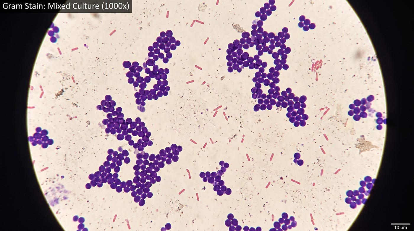
There’s a moment in every microbiology lab when something genuinely surprising happens. You’ve been going through steps — adding dyes, rinsing, waiting — and then you hold your slide up to the microscope and see two completely different colors where, just minutes ago, there was nothing visible at all. Some bacteria are deep purple. Others are pink.
That’s Gram staining. And what it’s actually showing you is one of the most fundamental differences between types of bacteria: the structure of their cell walls.
A Little History First
The Gram stain was developed in 1884 by a Danish microbiologist named Hans Christian Gram. What’s remarkable is that the technique he created has remained essentially unchanged ever since. It’s still one of the first things done when identifying an unknown bacterium — over 140 years later.
The reason it’s lasted so long is that it reveals something genuinely meaningful. Gram staining doesn’t just color bacteria arbitrarily. It sorts them into two categories — gram-positive and gram-negative — that correspond to real, fundamental differences in how their cell walls are built. And those differences affect everything from how bacteria behave, to which antibiotics can kill them.
What You Need for the Procedure
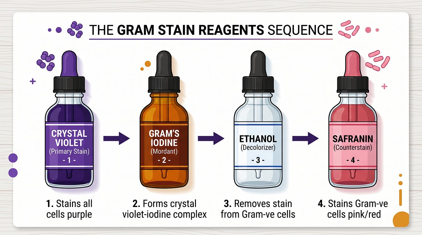
The Gram stain uses four reagents, each with a specific role:
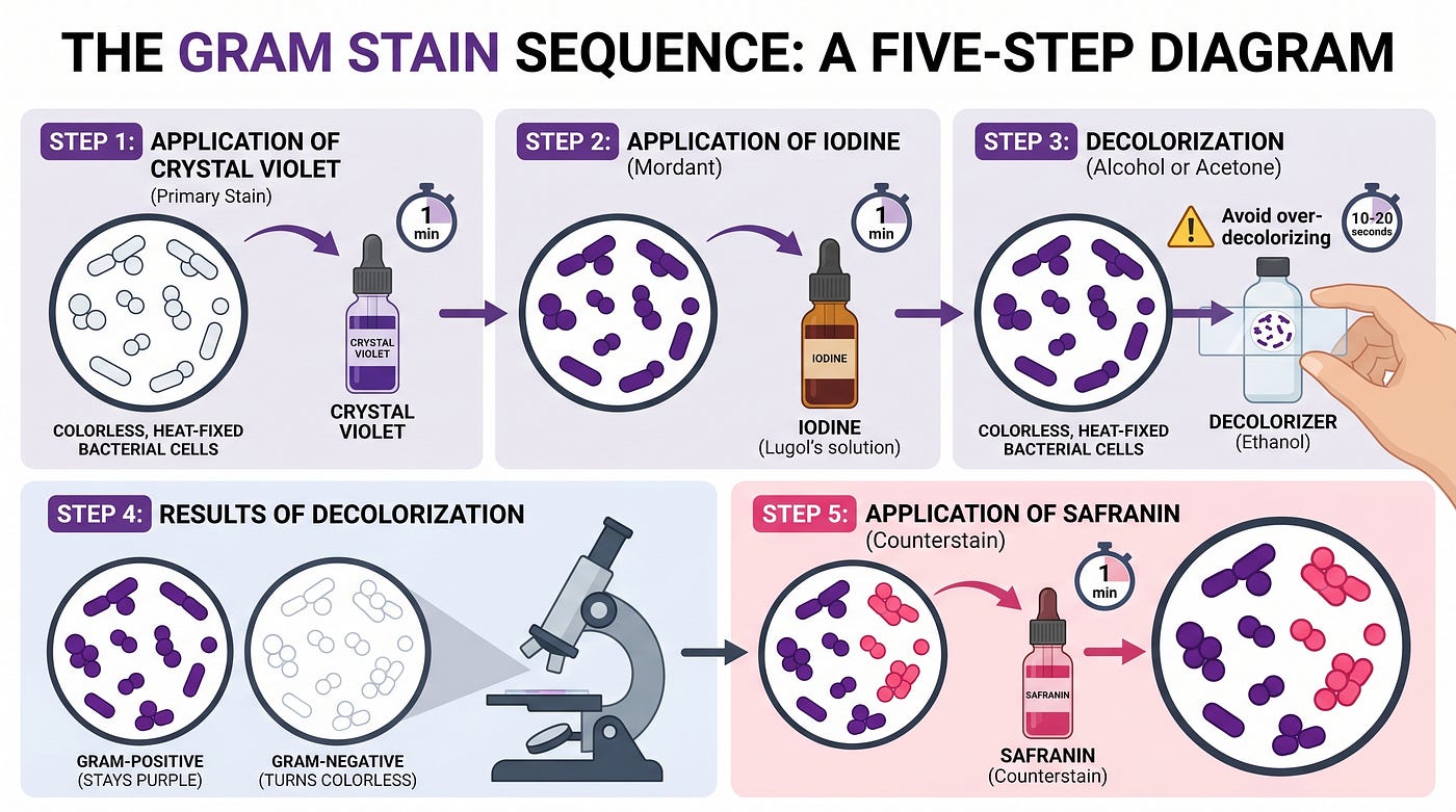
Crystal violet is the primary stain. It’s the purple dye that goes on first and temporarily colors all bacteria.
Iodine solution acts as a mordant — a fixing agent. It forms a complex with crystal violet and helps lock the dye into the cell wall. After iodine, both gram-positive and gram-negative bacteria are still purple.
Ethanol or acetone is the decolorizer. This is the step that separates the two groups. It washes the dye out of some bacteria but not others, depending on their cell wall thickness.
Safranin is the counterstain — a secondary pink/red dye added at the end. It stains the bacteria that lost their color during decolorization, making them visible again.
Step-by-Step: How the Procedure Works
Here’s the full sequence, from bacterial culture to microscope view.
1. Prepare the smear. Place a drop of fresh bacterial culture in the center of a clean glass slide using a dropper.
2. Heat-fix the sample. Pass the slide carefully through a Bunsen burner flame three times. This kills the bacteria and attaches them to the slide so they don’t wash off during staining.
3. Apply crystal violet (30–40 seconds). Flood the slide with the primary stain and let it sit. At this point, all bacteria on the slide turn purple.
4. Rinse gently with water (max 5 seconds). A short rinse removes unbound dye without disturbing what’s been taken up by the cells.
5. Apply iodine solution (1 minute). The iodine forms a crystal violet–iodine complex inside the cells, effectively trapping the dye. Both gram-positive and gram-negative bacteria are still purple here.
6. Rinse with water briefly (2 seconds).
7. Decolorize with ethanol or acetone. This is the critical step. The decolorizer is applied and then rinsed off. In gram-positive bacteria, the thick peptidoglycan wall dehydrates and tightens, trapping the crystal violet–iodine complex inside. The purple stays. In gram-negative bacteria, the thinner wall can’t hold the complex — the dye washes out and the cells become colorless again.
8. Apply safranin (20–30 seconds). The counterstain colors the now-colorless gram-negative bacteria pink or red. Gram-positive bacteria, still carrying their purple dye, are barely affected by the safranin.
9. Rinse with distilled water, dry with filter paper.
10. Add a cover slip and view under the compound microscope.
What You See (and Why)
After the procedure, gram-positive bacteria appear blue-purple and gram-negative bacteria appear pink-red. Here’s a summary of what happens at each step:
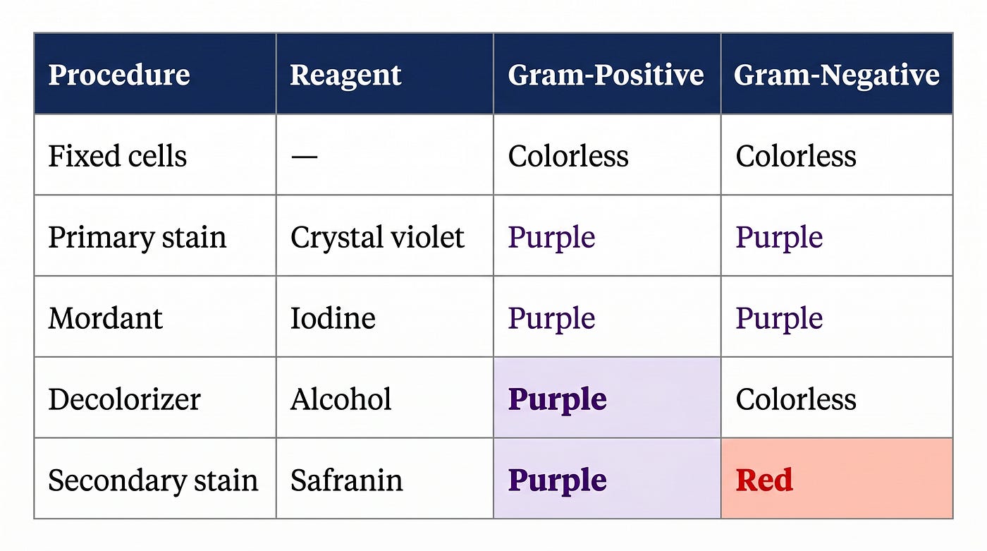
The difference only shows up at the decolorization step — and that difference is entirely explained by cell wall structure.
The Real Reason: Cell Wall Structure
This is where the biology gets interesting.
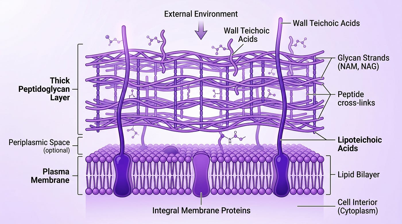
Gram-positive bacteria have a thick, dense layer of peptidoglycan sitting just outside their plasma membrane. Teichoic and lipoteichoic acids project outward from this layer. The overall negative charge of the wall helps the basic (positively charged) crystal violet dye bind strongly in the first place.
When the alcohol decolorizer is applied, it dehydrates the peptidoglycan, causing it to shrink and tighten. The spaces in the cell wall close up, physically trapping the crystal violet–iodine complex inside. It can’t escape. The cell stays purple.
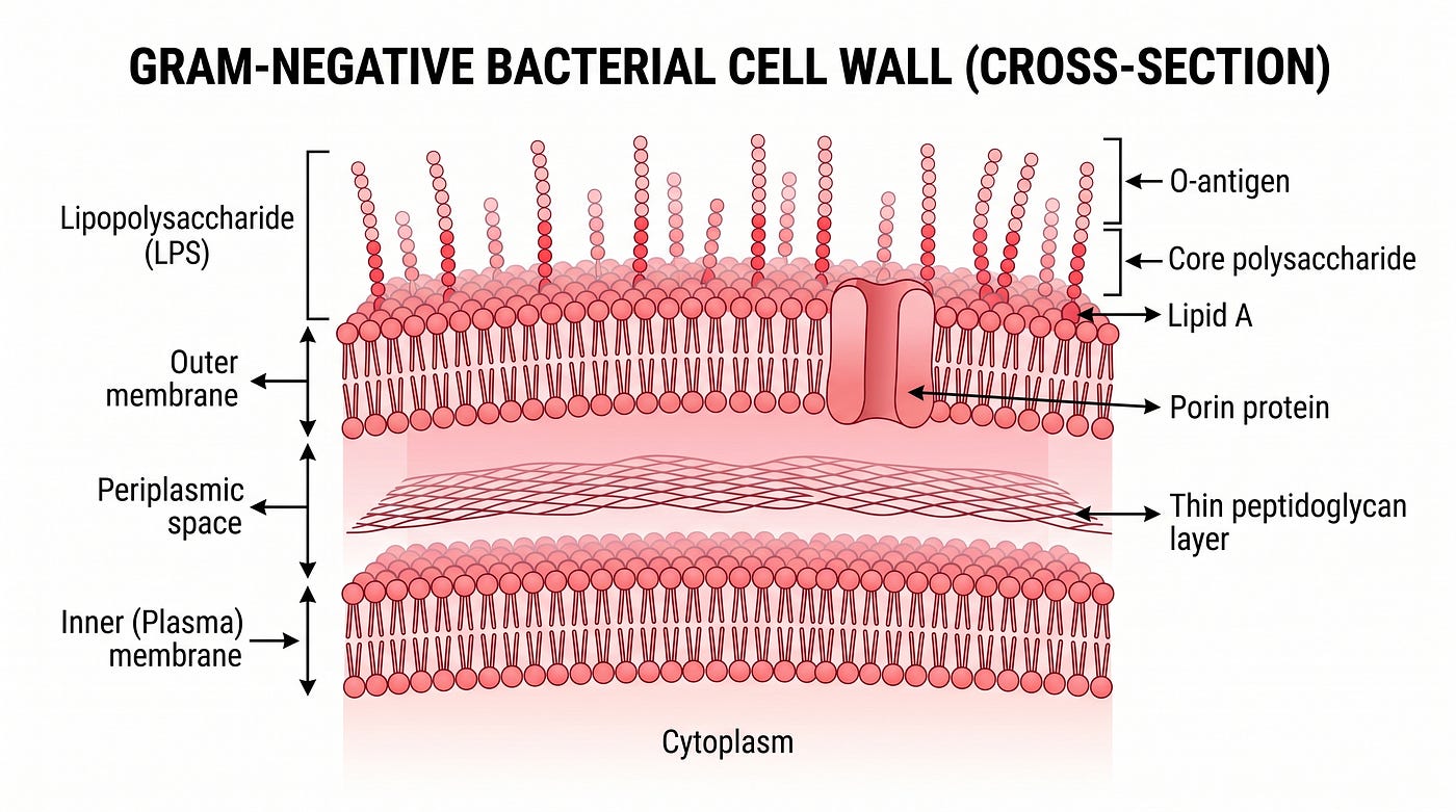
Gram-negative bacteria have a more complex, layered wall structure. There’s a thin peptidoglycan layer, but it’s sandwiched between the plasma membrane and an additional outer membrane. The gap between them is called the periplasmic space. The outer membrane contains lipopolysaccharide (LPS) and porin proteins that allow small molecules to pass through.
When the decolorizer is applied, it dissolves the lipopolysaccharide in the outer membrane, damaging the cell wall. The crystal violet–iodine complex, no longer contained, leaks out through the now-permeable wall. The cell becomes colorless — until safranin stains it pink.
Why This Matters Beyond the Lab
Gram staining isn’t just a classroom exercise. Clinically, knowing whether a pathogen is gram-positive or gram-negative is one of the first steps in choosing the right antibiotic. Many antibiotics target the cell wall — and because gram-positive and gram-negative walls are structurally so different, they’re vulnerable to different drugs.
For example, penicillin and related antibiotics work by disrupting peptidoglycan synthesis. They’re generally more effective against gram-positive bacteria, whose thick peptidoglycan is the dominant feature. Gram-negative bacteria, protected by their additional outer membrane, are often harder to target.
That’s why a quick Gram stain — which takes less than 15 minutes — can have real consequences for patient care.
Wrapping Up
The Gram stain sorts bacteria into two groups based on how their cell walls respond to a sequence of dyes and a decolorizing agent. Gram-positive bacteria hold onto crystal violet and appear purple. Gram-negative bacteria lose it and pick up safranin instead, appearing pink.
The deeper reason is purely structural: thick peptidoglycan traps the dye, thin peptidoglycan with an outer membrane doesn’t. A technique invented in 1884 is still doing its job, and understanding why it works makes it a lot easier to remember how it works.
메타데이터
- post_id
- dec34bf3c480
- slug
- why-some-bacteria-turn-purple-and-others-turn-pink-the-science-of-gram-staining-dec34bf3c480
- url
- https://medium.com/@milkyway.9628/why-some-bacteria-turn-purple-and-others-turn-pink-the-science-of-gram-staining-dec34bf3c480
- canonical_url
- https://medium.com/@milkyway.9628/why-some-bacteria-turn-purple-and-others-turn-pink-the-science-of-gram-staining-dec34bf3c480
- author_url
- https://medium.com/@milkyway.9628
- status
- ok
- fetched_at
- 2026-06-15 20:49:13