How proteins work together to support our vision
SBGrid member Antonia Roll-Mecak analyzes the structure of the RPGR-TTLL5 complex to understand the pathomechanism of retititis pigmentosa.
Publication Highlight
How proteins work together to support our vision
This publication highlight is part of the SBGrid Communities Project focused on science education and demonstrating how structural biology and preclinical science connect to medicine, a collaboration between SBGrid PI Piotr Sliz and Jamaine Davis of Belmont University.
Retinitis pigmentosa is a disease of the eye characterized by progressive degeneration of the retina. Photoreceptors within the retina are responsible for converting light into electrical signals that allow the brain to interpret them as an image. As these photoreceptors degenerate, affected individuals may experience a spectrum of visual impairment, which may present as visual field reduction, reduced night vision, and total blindness in severe cases.
X-linked retinitis pigmentosa (XLRP) is a rare, severe inherited retinal disease affecting an estimated 40,000 people globally. Seventy percent of X-linked retinitis pigmentosa cases are due to mutations in the retinitis pigmentosa GTPase regulator (RPGR), a multi-domain protein involved in normal retinal function and aging. Of these cases, 80 percent of the RPGR mutations causing retinitis pigmentosa are localized to the C-terminal binding domain (BD) of the protein. RPGR activation requires a member of the tubulin tyrosine ligase-like enzyme family, TTLL5, to add glutamate chains to RPGR in a process termed glutamylation. This interaction between the TTLL5 and its non-tubulin substrate RPGR is crucial for retinal health. Consequently, mutations in either protein are implicated in the development of X-linked retinitis pigmentosa and severe retinal degeneration.
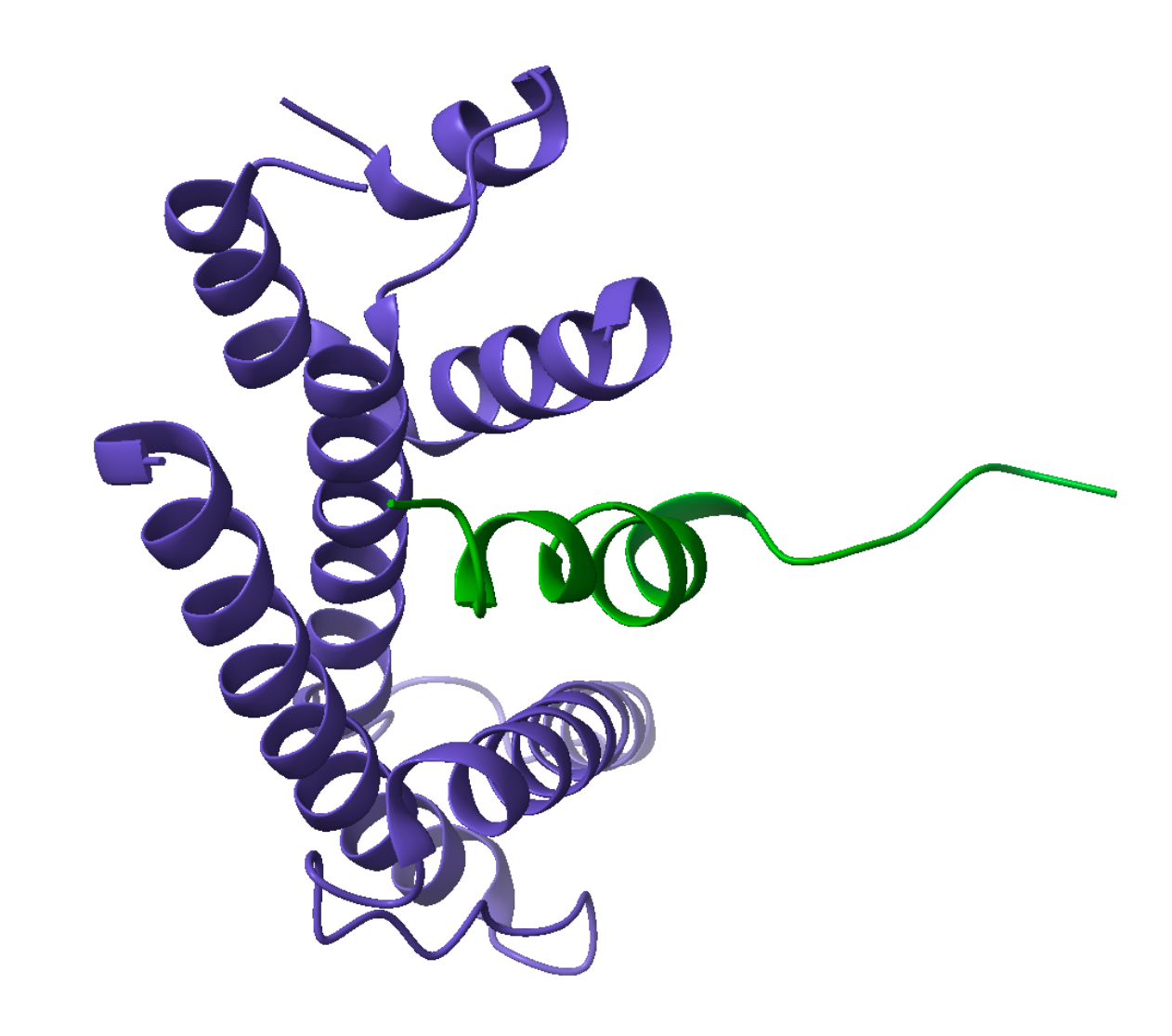
Structure of the RPGR BD–TTLL5 CID complex. RPGR BD in green. TTLL5 CID in purple. PDB 9PHH. CC by SBGrid.
SBGrid member Antonia Roll-Mecak and colleagues at the National Institutes of Health explored the structure of the RPGR C-terminal helix bound to the TTLL5 coactivator interacting domain (CID), an interaction needed for glutamylation. They utilized X-ray crystallization to determine the structure of the RPGR-TTLL5 complex at the atomic level. This work contributes to a growing body of research focused on proteins involved in the development of retinitis pigmentosa and macular degeneration of unknown etiology.
The researchers found key aromatic amino acid residues on the RPGR BD and TTLL5 CID within their binding interface. Point mutations of tryptophan at position 1141 and leucine at position 1151 on RPGR as well as phenylalanine at positions 680 and 755 on TTLL5 disrupt the RPGR–TTLL5 interaction and are associated with retinal disease. The authors also resolve an X-ray crystal structure of the RPGR BD–TTLL5 CID complex to 2.8 Å, disclosing a short helix with aromatic amino acids on RPGR that sits in a binding pocket created by an 𝛼-helical bundle of TTLL5.
This study reveals that mutations of aromatic amino acids on either RPGR or TTLL5 prevent glutamylation, revealing that the molecular mechanism, particularly between the CID of this glutamylase and its associated non-tubulin substrate underlies the primary cause of retinitis pigmentosa. Further understanding of this mechanism may inform diagnostic and treatment strategies to improve outcomes for those with RPGR mutation-dependent retinal disease.
Read more in ***Journal of Cell Biology***.
By Mudiare Ikoba, Meharry Medical College (Mudia Ikoba)
Mudiare “Mudia” Ikoba is a second-year medical student at Meharry Medical College. She completed her B.A. in Biology with a Biochemistry Concentration and a minor in Social Inequalities at Dartmouth College. When she is not studying, she enjoys being outdoors, exercising, cooking, and spending time with family and friends.
메타데이터
- post_id
- e4e1a30a6850
- slug
- how-proteins-work-together-to-support-our-vision-e4e1a30a6850
- url
- https://medium.com/sbgrid-community-news/how-proteins-work-together-to-support-our-vision-e4e1a30a6850
- canonical_url
- https://medium.com/sbgrid-community-news/how-proteins-work-together-to-support-our-vision-e4e1a30a6850
- author_url
- https://medium.com/@sbgrid
- status
- ok
- fetched_at
- 2026-06-09 15:37:30