A Validated Endotoxin Detection Workflow for Next-Generation LNP-Delivered mRNA Vaccines
Article Title:
A Validated Endotoxin Detection Workflow for Next-Generation LNP-Delivered mRNA Vaccines
Article Title:
Study on Bacterial Endotoxin Detection Methods for mRNA Vaccines Based on Lipid Nanoparticle Delivery Systems
Published in:
Chinese Journal of Microbiology and Immunology, November 2025
Author Affiliations:
Respiratory Virus Vaccine Division, National Institutes for Food and Drug Control (NIFDC) Zhuhai Lifanda Biotechnology Co., Ltd.

Published Article
Bacterial Endotoxin Detection Methods Using Recombinant Factor C (rFC) for LNP-mRNA Vaccines
Using the gel-clot limit test described in Part III of the 2020 Chinese Pharmacopoeia, the authors evaluated HSV-2, RSV, and VZV mRNA vaccines. Through optimization of sample dilution factors, diluents, and interference testing, suitable conditions for bacterial endotoxin detection were established.
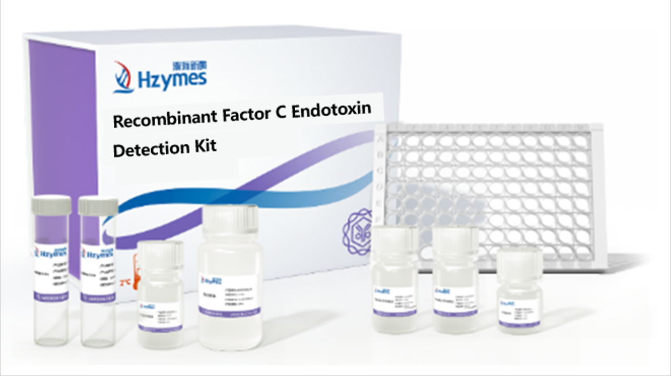
In this study, Hzymes’ Recombinant Factor C (rFC) Endotoxin Assay Kit was used to validate the detection results.
Lipid Nanoparticle (LNP) Structure and Its Impact on mRNA Vaccine Endotoxin Testing
mRNA technology, known for rapid design, high production efficiency, and strong immunogenicity, has opened new avenues in vaccines, oncology, infectious diseases, and autoimmune disorders. The emergency use of COVID-19 mRNA vaccines accelerated its clinical validation and industrialization.
mRNA is delivered into the body via lipid nanoparticles (LNPs), which typically contain cationic lipids, cholesterol, phospholipids, and PEGylated lipids. These components encapsulate nucleic acids through electrostatic interactions.

Structural Diagram of LNP-mRNA
LNP-mRNA vaccines are primarily formulated as injectables, and bacterial endotoxin testing is a critical quality-control requirement. Endotoxins, structural components of Gram-negative bacterial cell walls, consist of O-antigen, core polysaccharide, and lipid A. Even trace amounts (1–5 ng/kg body weight) can trigger severe adverse reactions such as fever, sepsis, DIC, or shock.
Since LNPs are formed by hydrophobic lipid self-assembly, endotoxins may bind to lipid components during detection via electrostatic or hydrophobic interactions. Studies have shown that endotoxins can be adsorbed by liposomes when tested with LAL reagents, causing interference. Diluting lipid concentrations can reduce endotoxin–lipid interactions, and adding divalent cations like magnesium may also suppress interference.

Structural Diagram of Endotoxin
Materials and rFC Testing Methods for Accurate Endotoxin Analysis in LNP-mRNA Formulations
A portion of the study utilized different batches of Hzymes’ rFC assay kits along with unmasking buffer and optimized dilution factors to eliminate LNP-related interference, establish an endotoxin detection method suitable for LNP-mRNA vaccines, and validate the results.
Test Samples
HSV-2, RSV, and VZV mRNA vaccines provided by Zhuhai Lifanda Biotechnology Co., Ltd.
Reagents
Recombinant Factor C assay kits (range: 0.005–5 EU/mL; batch numbers HH20250401R, HH20250101R, HH20240601R) provided by Wuhan Hzymes Biotech Co., Ltd.
Instrument
SpectraMax i3x fluorescence microplate reader.
Detection Procedures
-
Using the formula MVD = cL/λ (c = 1.0 mL/mL; L = 30 EU/mL; λ = 0.005 EU/mL), the maximum valid dilution (MVD) was calculated as 6000×. Based on preliminary data, samples were diluted 100× using a Ca/Mg unmasking buffer.
-
Following the kit instructions, endotoxin standards were prepared at 5, 0.5, 0.05, and 0.005 EU/mL, with endotoxin-free water as the negative control. Each standard, sample, and control (100 μL) was added to 96-well plates in duplicate, followed by 100 μL of substrate. The instrument was set to Ex 380 nm / Em 440 nm. Fluorescence was recorded at 0 h and after a 1 h incubation at 37°C. ΔRFU was calculated by subtracting initial fluorescence, and net ΔRFU was obtained by subtracting the negative control.
-
Data Analysis: A linear fit was generated using log concentration (x-axis) and log net ΔRFU (y-axis). Spike recoveries were calculated as: Recovery (%) = (C_spiked — C_sample) / 0.5 EU/mL × 100% A recovery of 50–200% indicated no interference.

Validation
1. Standard Curve Validation:
Three batches of rFC kits produced standard curves with R² = 0.999, 0.990, and 0.999, all ≥0.980, meeting Chinese Pharmacopoeia 2020 requirements. Negative controls were below the lowest standard point (0.005 EU/mL). These curves were therefore valid for endotoxin quantification.
2. Spike Recovery and Sample Testing:
Three batches each of HSV-2, RSV, and VZV vaccines were diluted 100×. One set served as the sample group; the other was spiked to 0.5 EU/mL. Endotoxin concentrations and spike recoveries were calculated using the standard curve equations. Recovery within 50–200% indicated no interference. Qualified samples were then used to calculate final endotoxin content based on the dilution factor.
Endotoxin Testing Results: rFC Assay Performance, Recovery Rates, and LNP Interference Removal
Standard Curve Reliability:
The three kit batches yielded R² values of 0.999, 0.990, and 0.999, all exceeding 0.980. Negative controls remained below the lowest standard point, meeting the validity requirements of General Chapter 1143 (photometric method) in the 2020 Chinese Pharmacopoeia.

Spike Recovery and Detection Results:
Across HSV-2, RSV, and VZV vaccine samples, rFC-based endotoxin measurements showed spike recoveries between 79.5% and 102.4%, within the acceptable 50–200% range. Endotoxin levels were <30 EU/mL and consistent with results obtained by the gel-clot method.

Conclusion and Future Outlook for rFC Endotoxin Assays in LNP-mRNA Vaccine Quality Control
LNP samples may interfere with LAL-based endotoxin assays. Using the unmasking buffer in the rFC kit effectively eliminated this interference. The rFC method produced results consistent with the gel-clot method, supporting its reliability.
This study provides an effective strategy for establishing endotoxin testing methods for LNP-mRNA vaccines, ensuring accuracy by minimizing interference. With the advancement of endotoxin detection technologies, methods such as rFC offer strong complementary value to traditional LAL assays, providing more comprehensive quality-control capabilities for LNP-based mRNA vaccines.
Recombinant Factor C (rFC) Endotoxin Detection Kit — Ordering Information

To obtain the original PDF, please contact: https://www.hzymesbiotech.com/index/user/register?url=/ Please verify: Recombinant Factor C
메타데이터
- post_id
- e75f9f77eac5
- slug
- a-validated-endotoxin-detection-workflow-for-next-generation-lnp-delivered-mrna-vaccines-e75f9f77eac5
- url
- https://medium.com/@hzymesmarketing/a-validated-endotoxin-detection-workflow-for-next-generation-lnp-delivered-mrna-vaccines-e75f9f77eac5
- canonical_url
- https://medium.com/@hzymesmarketing/a-validated-endotoxin-detection-workflow-for-next-generation-lnp-delivered-mrna-vaccines-e75f9f77eac5
- author_url
- https://medium.com/@hzymesmarketing
- status
- ok
- fetched_at
- 2026-06-09 15:37:30