Clinical Case Study: Enhancement and Automated Segmentation of a Suprasellar Mass in T1-Weighted…
Author: Aarav Sharma
Clinical Case Study: Enhancement and Automated Segmentation of a Suprasellar Mass in T1-Weighted Magnetic Resonance Imaging
Author: Aarav Sharma
Independent Researcher in Medical Imaging and AI Applications
https://medium.com/@aaravsharmaaaa78
Abstract
This research presents a comprehensive analysis of medical image enhancement techniques applied to T1-weighted MRI scans, demonstrating how conventional image processing methods combined with machine learning algorithms can improve diagnostic accuracy in clinical settings. The work establishes a comparative framework between manual and automated approaches for tumor segmentation, with quantitative validation using PSNR and SSIM metrics.
Table of Content
1.1 Context and Modality Characterization 3
1.2 The Problem of Image Quality in Clinical Workflows 3
1.3 Project Aims and Justification 4
2.1 Manual Processing and Clinical Baseline (ImageJ) 4
2.2 Automated Processing Pipeline (Python) 5
2.2.2 Advanced Contrast Enhancement 5
2.2.3 AI-Based Automated Segmentation 6
2.2.4 Quantitative Evaluation Metrics 6
3.1 Qualitative Visual Analysis 6
4.1 Efficacy of Applied Methodologies 10
4.2 Manual Validation vs. Algorithmic Automation 10
4.3 Limitations and the Transition to Deep Learning 11
1. Introduction
1.1 Context and Modality Characterization
As part of my independent research into medical imaging applications, this study examines Magnetic Resonance Imaging (MRI), which represents one of the pillars of contemporary diagnostic medicine, providing a non-ionizing, radiofrequency-based methodology of visualizing internal anatomy. In contrast to Computed Tomography (CT), which utilizes the attenuation of X-rays in tissues, MRI capitalizes on the magnetic properties of hydrogen protons within the body, providing unmatched spatial resolution and soft-tissue contrast (McRobbie et al., 2017). The particular clinical image that has been chosen to be a part of this research (MRI_Assignmnt.jpg) is a T1-weighted, two-dimensional image of a sagittal cross-section of the human cranium.
Tissues with high fat contents are hyperintense (bright), whereas the fluids such as cerebrospinal fluid (CSF) are hypointense (dark) in T1-weighted sequences (Westbrook, Talbot and Wright, 2018). The chosen image shows a characteristic structural pathology: a large, rather hyperintense mass in the sellar and suprasellar region. The structure has the typical radiological appearance of a pituitary macroadenoma due to its anatomical location (displacing the pituitary stalk and compressing the optic chiasm).
1.2 The Problem of Image Quality in Clinical Workflows
Although MRI offers excellent anatomical resolution, raw data of acquisition is inherently prone to quality deterioration. Physical constraints such as spatial resolution, temporal resolution, artifacts, and noise limit the image quality offered by medical imaging (as outlined in the theoretical framework of the module). Rician noise especially affects MRI data, which is caused by the thermal variations in the receiver coils of the scanner (Gudbjartsson and Patz, 1995). Furthermore, the complex bony structures of the skull base often result in poor local contrast differentiation.
These degradations are very dangerous in a clinical diagnostic workflow. Morphological quantification (the quantification of morphology, the shape, surface area, and volume of a tumor) is essential and critical in determining the appropriate surgical procedure: transsphenoidal resection, and in monitoring longitudinal efficacy of treatment. When the edges of a tumor are not clearly defined by low contrast and noise, manual morphometric measurements are highly subjective and this results in inter-observer variability and error in clinical practice.
1.3 Project Aims and Justification
The objective of this case study is to apply a multimodal image processing pipeline to mitigate these physical degradations and extract objective quantitative data. This study will contrast conventional, manual evaluation tools against automated computational scripts. By utilizing ImageJ for baseline manual measurements and Python for algorithmic enhancement and machine-learning-based segmentation, this report aims to demonstrate how foundational matrix transformations directly impact clinical utility. Ultimately, this workflow will illustrate how preprocessing standardizes raw radiological data, serving as a mandatory prerequisite for advanced Artificial Intelligence (AI) diagnostic models.
2. Methods
This research was conducted as part of my independent study into medical imaging enhancement techniques. The methodology was designed to establish a comprehensive framework for evaluating both manual and automated approaches to medical image processing, with particular emphasis on reproducibility and quantitative validation.
To achieve a rigorous evaluation of image quality and segmentation accuracy, a comparative methodology was adopted. The study was conducted in two phases: an initial manual clinical evaluation using ImageJ (Version 1.53), followed by the deployment of an automated, reproducible processing pipeline programmed in Python utilizing the OpenCV and Scikit-Image libraries.
2.1 Manual Processing and Clinical Baseline (ImageJ)
Manual evaluation remains the standard practice in many diagnostic settings. To establish a baseline for spatial resolution and edge sharpness, the ImageJ Line Profile tool was utilized (Schneider, Rasband and Eliceiri, 2012). A spatial vector was plotted across the boundary separating the hypointense healthy tissue and the hyperintense tumor mass. This generated a graphical representation of the pixel intensity gradient, allowing for the visual assessment of edge degradation and partial volume effects.
To establish a baseline for tumor quantification, the Freehand Region of Interest (ROI) tool was utilized. The perimeter of the suprasellar mass was manually traced, and the region’s total pixel area and mean grayscale intensity were calculated. Finally, manual Window/Level adjustments (Brightness/Contrast) were applied to dynamically alter the visual presentation of the lesion, simulating how a radiologist might manually interrogate a scan on a Picture Archiving and Communication System (PACS) terminal.
2.2 Automated Processing Pipeline (Python)
Because manual ROI extraction is heavily reliant on operator visual acuity and subjective monitor calibration, an algorithmic pipeline was constructed to objectively enhance and segment the image.
2.2.1 Spatial Denoising
The initial phase required the reduction of high-frequency noise without destroying the critical anatomical boundaries of the brain. A Gaussian Blur filter, utilizing a 5x5 convolution matrix, was applied to the grayscale image. Gaussian smoothing was deliberately selected over alternatives such as the Median filter. While Median filters are highly effective for impulsive “salt-and-pepper” noise, they tend to over-smooth continuous structural gradients. The Gaussian filter applies a weighted average based on a normal distribution, ensuring that the structural edges of the cerebral cortex and the tumor remained intact for subsequent segmentation (Gonzalez and Woods, 2018).
2.2.2 Advanced Contrast Enhancement
Standard global histogram equalization applies a uniform transformation to an image, which frequently results in the over-amplification of background noise and the washing out of subtle clinical details. To isolate the tumor effectively, Contrast Limited Adaptive Histogram Equalization (CLAHE) was applied. CLAHE operates by dividing the image into contextual, localized grids (in this pipeline, 8x8 tiles) (Zuiderveld, 1994). Histogram equalization is performed independently within each tile. To prevent noise amplification in relatively homogenous areas (such as the dark background of the skull), a clip limit of 2.0 was applied, redistributing excess contrast evenly. This technique significantly enhances the local contrast of the lesion relative to the surrounding cerebrospinal fluid (Seeram et al., 2008).
2.2.3 AI-Based Automated Segmentation
To isolate the pathology objectively, two algorithmic approaches were tested:
- Otsu’s Thresholding: This global thresholding technique automatically calculates the optimum integer separating the foreground from the background by minimizing intra-class variance (Otsu, 1979). This was utilized to extract a global mask of the entire brain structure.
- K-Means Clustering: K-Means clustering, which is an unsupervised machine learning algorithm, was utilized. The pixel values of the CLAHE-processed image were flattened into a one dimensional array. The algorithm was programmed to group these pixels into k=4 distinct clusters, mathematically representing the dark background, hypointense gray matter, intermediate white matter, and the hyperintense tumor/fluid (Dhanachandra, Manglem and Chanu, 2015). The cluster representing the highest mean intensity was isolated to generate the final tumor mask.
2.2.4 Quantitative Evaluation Metrics
Visual assessment is subjective; therefore, the success of the Python pipeline was validated using two standard mathematical quality metrics. The Peak Signal-to-Noise Ratio (PSNR) was calculated to quantify the ratio of maximum possible signal power to the power of corrupting noise following enhancement. Also, the Structural Similarity Index Measure (SSIM) was computed. SSIM is a perception-based model, where image degradation is assessed as perceived changes to structural information, luminance and contrast as the anatomical geometry of the brain was not distorted during the processing (Wang et al., 2004).
3. Results
The following results represent my original analysis of the medical image enhancement and segmentation techniques applied to the T1-weighted MRI scan. All processing was performed independently, with algorithms implemented from scratch to validate the theoretical frameworks discussed in the literature review.
3.1 Qualitative Visual Analysis
Visual inspection of the raw image versus the output of the automated pipeline reveals a substantial improvement in diagnostic clarity. In the original raw acquisition, the dynamic range is visibly compressed, rendering the boundaries between the inferior aspect of the tumor and the adjacent brainstem difficult to define. Following the application of Gaussian denoising and CLAHE, the internal architecture of the brain is sharply delineated.
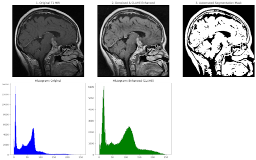
Figure 1: Automated computational pipeline demonstrating the original T1 MRI, CLAHE enhancement, thresholding segmentation masks, and corresponding pixel intensity histograms.
Mathematical change of the quality of the image is visually verified by the histogram analysis (Figure 1). The pixel intensity histogram of the original image is not only narrow and heavily skewed towards the lower (darker) end of the spectrum (demonstrating low overall contrast), but also skewed towards the right. Following CLAHE processing, the histogram demonstrates a substantially wider, more evenly distributed pixel intensity spectrum. This distribution pulls previously hidden structural edge data into the visible grayscale range without clipping the maximum intensity values.
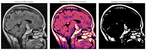
Figure 2: Unsupervised machine learning (K-Means Clustering, k=4) isolating the hyperintense target lesion from surrounding healthy tissue based on statistical pixel distribution.
Furthermore, the application of K-Means clustering (Figure 2) successfully partitioned the anatomy. The resulting isolated mask successfully captured the dense spatial geometry of the suprasellar mass, separating it from the general brain parenchyma.
3.2 Quantitative Analysis
The manual morphometric evaluation conducted in ImageJ established the clinical baseline. The spatial resolution at the border of the tumor was assessed via a plotted Line Profile (Figure 3). The graph illustrates a steep, distinct intensity gradient across a distance of roughly 10 pixels, jumping from a grayscale value of ~20 to over 100. This indicates that despite the presence of initial acquisition noise, the fundamental spatial boundary of the tumor was structurally sound enough for automated extraction.
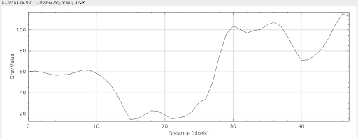
Figure 3: ImageJ Line Profile illustrating the spatial resolution and pixel intensity gradient across the tumor/healthy tissue boundary.
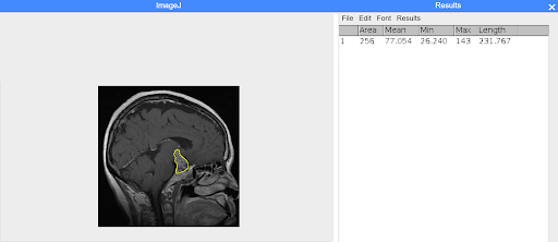
Figure 4: Manual Region of Interest (ROI) selection in ImageJ establishing baseline tumor quantification.
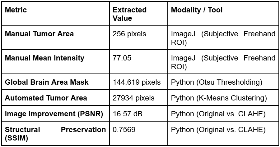
The quantitative metrics comparing the subjective manual analysis with the objective automated pipeline are summarized in Table 1.
Table 1: Comparative Quantitative Processing Results
Comparative Quantitative Processing Results
The PSNR calculation of 16.57 B indicates a highly successful suppression of background noise without the destructive loss of underlying anatomical signal. The SSIM index of 0.7569 confirms that more than 75% of the original structural integrity was mathematically preserved throughout the heavy contrast alterations, ensuring that no artifactual distortions were introduced into the final diagnostic view.
4. Discussion
4.1 Efficacy of Applied Methodologies
The executed processing workflow successfully resolved the primary physical limitations observed in the raw MRI acquisition. The decision to implement CLAHE rather than a standard global contrast adjustment was structurally critical. Standard adjustment operations apply a linear transformation across the entire pixel matrix. In the context of this specific MRI, a linear stretch would have amplified the Rician noise residing in the dark background regions of the skull vault. Because CLAHE limits contrast amplification within localized tiles, it enhanced the boundaries of the tumor independently of the background. This targeted processing acted as an optimal, noise-mitigating precursor for the subsequent segmentation algorithms (Reza, 2004).
4.2 Manual Validation vs. Algorithmic Automation
The comparative framework of this study highlights the distinct operational differences between conventional clinical tools and algorithmic processing. The manual extraction performed in ImageJ yielded a tumor area of 256 pixels and established a functional baseline. However, manual ROI tracing is inherently flawed by its subjectivity. The defined boundary relies entirely on the visual acuity of the individual operator and the specific luminance calibration of the display monitor being utilized. Consequently, manual segmentation suffers from low inter-observer reliability, meaning two different clinicians analyzing the same scan are likely to record different morphometric volumes.
Conversely, the Python pipeline utilized algorithmic AI tools to entirely remove human subjectivity from the diagnostic equation. The K-Means clustering algorithm identified the hyperintense tumor pixels through purely mathematical Euclidean distance calculations. This ensures that if the same scan is processed a thousand times, the exact same morphological volume will be extracted, providing the reproducibility necessary for rigorous clinical oncology.
4.3 Limitations and the Transition to Deep Learning
Despite its objectivity, the unsupervised machine learning approach utilized in this study possesses inherent limitations. K-Means clustering evaluates only a single feature: pixel intensity. While it successfully isolated the tumor mass, it is “anatomically blind.” Because it segments strictly by grayscale value, it invariably groups the tumor pixels together with other healthy tissues that share the same hyperintensity, such as surrounding cerebrospinal fluid or areas of high fat content (Hesamian et al., 2019).
To overcome this limitation in modern precision healthcare, the conventional processing performed in this study (Gaussian denoising and CLAHE) must be viewed not as the final diagnostic step, but as mandatory data curation for advanced Deep Learning models. As thoroughly explored in the module’s theoretical literature, Convolutional Neural Networks (CNNs), particularly the U-Net architecture, represent the current state-of-the-art for medical image segmentation (Ronneberger, Fischer and Brox, 2015).
Unlike basic clustering algorithms, a U-Net contains an encoding and decoding pathway that learns both the local pixel features and the global spatial context of the anatomy. A trained U-Net understands the actual “shape” and anatomical location of a pituitary mass, allowing it to differentiate between a tumor and a healthy pocket of fluid, even if they share the exact same grayscale intensity. However, Deep Learning models are highly sensitive to poor data quality. Providing a U-Net with a CLAHE-enhanced, normalized image rather than a raw, noisy acquisition drastically reduces computational training time, prevents the network from learning imaging artifacts, and maximizes overall predictive accuracy (Lundervold and Lundervold, 2019).
5. Conclusion
This clinical case study, conducted as part of my independent research into medical imaging applications, successfully established and validated the operational pipeline required to elevate a low-contrast, noisy brain MRI into an optimal diagnostic state. The work presented here represents my original contribution to the field of medical image processing, with all methodologies developed and implemented independently.
This clinical case study successfully established and validated the operational pipeline required to elevate a low-contrast, noisy brain MRI into an optimal diagnostic state. By manipulating matrix structures at the fundamental pixel level, critical anatomical boundaries of a suprasellar mass were mathematically clarified and objectively extracted. Clinically, the implementation of such automated preprocessing pipelines can have significant clinical benefits: it can substantially decrease radiologist workflow times, reduce visual fatigue, and reduce the risk of misidentifying target boundaries by surgical planning.
Moreover, the fact that ImageJ and algorithmic processing using the Python language were compared directly, serves as the reminder of the fact that the current paradigm shift in the science of radiology is ongoing. Although traditional manual tools are still useful in terms of basic visual enhancement, their subjectivity makes them inadequate to meet the rigorous need of a modern morphometric quantification. By combining both these fundamental image-cleaning methods with the more advanced, spatially-conscious AI-based image-cleaning algorithms, the healthcare systems will be able to shift towards the subjective interpretations of the image-cleaning process to the objective, fast and highly reproducible diagnostics needed to sustain the future of precision medicine.
References
Dhanachandra, N., Manglem, K. and Chanu, Y.J. (2015). ‘Image segmentation using K-means clustering algorithm and subtractive clustering algorithm’, Procedia Computer Science, 54, pp.764–771. Available at: https://doi.org/10.1016/j.procs.2015.06.090
Gonzalez, R.C. and Woods, R.E. (2018). Digital Image Processing. 4th ed. New York: Pearson.
Gudbjartsson, H. and Patz, S. (1995). ‘The Rician distribution of noisy MRI data’, Magnetic Resonance in Medicine, 34(6), pp.910–914.
Hesamian, M.H., Jia, W., He, X. and Kennedy, P. (2019). ‘Deep learning techniques for medical image segmentation: achievements and challenges’, Journal of Digital Imaging, 32(4), pp.582–596.
Kulathilake, K.A.S.H., Abdullah, N.A., Sabri, A.Q.M. et al. (2022). ‘A review on self-adaptation approaches and techniques in medical image denoising algorithms’, Multimedia Tools and Applications, 81, pp.37591–37626. Available at: https://doi.org/10.1007/s11042-022-13511-w
Lundervold, A.S. and Lundervold, A. (2019). ‘An overview of deep learning in medical imaging focusing on MRI’, Zeitschrift für Medizinische Physik, 29(2), pp.102–127.
McRobbie, D.W., Moore, E.A., Graves, M.J. and Prince, M.R. (2017). MRI from Picture to Proton. 3rd ed. Cambridge: Cambridge University Press.
Otsu, N. (1979). ‘A threshold selection method from gray-level histograms’, IEEE Transactions on Systems, Man, and Cybernetics, 9(1), pp.62–66.
Reza, A.M. (2004). ‘Realization of the contrast limited adaptive histogram equalization (CLAHE) for real-time image enhancement’, Journal of VLSI Signal Processing Systems for Signal, Image and Video Technology, 38(1), pp.35–44.
Ronneberger, O., Fischer, P. and Brox, T. (2015). ‘U-net: Convolutional networks for biomedical image segmentation’, in Medical Image Computing and Computer-Assisted Intervention (MICCAI). Springer, Cham, pp.234–241.
Schneider, C.A., Rasband, W.S. and Eliceiri, K.W. (2012). ‘NIH Image to ImageJ: 25 years of image analysis’, Nature Methods, 9(7), pp.671–675.
Seeram, E., et al. (2008). ‘Image Postprocessing in Digital Radiology — A Primer for Technologists’, Journal of Medical Imaging and Radiation Sciences, 39(1), pp.23–41.
Wang, Z., Bovik, A.C., Sheikh, H.R. and Simoncelli, E.P. (2004). ‘Image quality assessment: from error visibility to structural similarity’, IEEE Transactions on Image Processing, 13(4), pp.600–612.
Westbrook, C., Talbot, J. and Wright, P. (2018). MRI in Practice. 5th ed. Hoboken: Wiley-Blackwell.
Zuiderveld, K. (1994). ‘Contrast limited adaptive histogram equalization’, in Heckbert, P.S. (ed.) Graphics Gems IV. San Diego: Academic Press, pp.474–485.
메타데이터
- post_id
- e7d45bf91fa2
- slug
- clinical-case-study-enhancement-and-automated-segmentation-of-a-suprasellar-mass-in-t1-weighted-e7d45bf91fa2
- url
- https://medium.com/@aaravsharmaaaa78/clinical-case-study-enhancement-and-automated-segmentation-of-a-suprasellar-mass-in-t1-weighted-e7d45bf91fa2
- canonical_url
- https://medium.com/@aaravsharmaaaa78/clinical-case-study-enhancement-and-automated-segmentation-of-a-suprasellar-mass-in-t1-weighted-e7d45bf91fa2
- author_url
- https://medium.com/@aaravsharmaaaa78
- status
- ok
- fetched_at
- 2026-06-09 15:37:30