The Science Behind Gel Electrophoresis: How DNA and RNA Find Their Way
Gel electrophoresis is a fundamental technique used in molecular biology to separate nucleic acids (DNA and RNA) based on their size, shape…
The Science Behind Gel Electrophoresis: How DNA and RNA Find Their Way
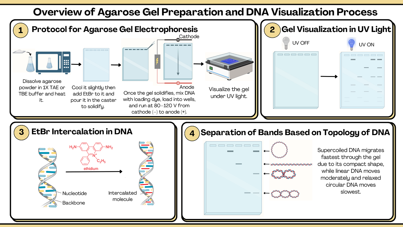
The figure illustrates process and visualization of agarose gel electrophoresis. Created using BioRender.com contents and book “Molecular Biology of the Gene”.
Gel electrophoresis is a fundamental technique used in molecular biology to separate nucleic acids (DNA and RNA) based on their size, shape and charge. DNA molecules carry a negative charge due to their phosphate backbone, so when an electric field is applied, they migrate toward the positive electrode (anode). The gel, usually made of agarose, a polysaccharide extracted from seaweed, acts like a molecular sieve that slows down larger fragments while allowing smaller ones to move more easily. As a result, small DNA fragments travel faster and farther, while larger fragments move more slowly and stay near the starting point. Over time, the fragments separate into distinct bands, each containing DNA molecules of similar size, resembling the steps of a ladder.
Visualizing DNA and Understanding Gel Types
To visualize the separated DNA, the gel is stained with ethidium bromide (EtBr), a flat, positively charged molecule that intercalates between the stacked bases of DNA. Under UV light, EtBr-bound DNA fluoresces, allowing the distinct bands to be seen clearly.
Different gels are used depending on the resolution needed:
- Agarose gels: suitable for larger DNA fragments (thousands of base pairs)
- Polyacrylamide gels: provide higher resolution for smaller fragments — capable of separating DNA differing by just a single base pair, though typically only up to about 1000 bases.
The shape or topology of DNA also affects its movement. Supercoiled DNA is more compact and moves fastest, linear DNA moves at a moderate rate, and circular (relaxed) DNA migrates the slowest, even though they are of equal size, because it encounters more resistance from the gel.
Protocol: Preparing and Running an Agarose Gel for DNA Electrophoresis
- Prepare the gel: Dissolve 1 g agarose in 100 mL 1× TAE/TBE buffer, heat until clear, cool slightly, then add ethidium bromide (0.5 µg/mL) and pour into a tray with a comb to solidify.
- Set up the tank: Place the gel in the electrophoresis chamber and cover it with 1× buffer.
- Load samples: Mix DNA with loading dye, pipette into wells, and include a DNA ladder for size reference.
- Run and visualize: Apply 80–120 V until separation occurs, then view fluorescent DNA bands under UV light.
Separation of RNA
RNA molecules, like DNA, are negatively charged, but being single-stranded, they often form complex secondary and tertiary structures that can alter their migration. To prevent this and ensure movement depends only on molecular weight, RNA is treated with glyoxal, which reacts with the amino groups in the bases and prevents base pairing. In this way, RNA too can be separated accurately by size.
Gel electrophoresis is not limited to nucleic acids, it also plays a vital role in protein analysis. In later discussions, we’ll explore how similar principles are applied to visualize and separate proteins, revealing their size, structure and purity.
메타데이터
- post_id
- f8f0b9edf8bb
- slug
- the-science-behind-gel-electrophoresis-how-dna-and-rna-find-their-way-f8f0b9edf8bb
- url
- https://medium.com/@palakparidhi123/the-science-behind-gel-electrophoresis-how-dna-and-rna-find-their-way-f8f0b9edf8bb
- canonical_url
- https://medium.com/@palakparidhi123/the-science-behind-gel-electrophoresis-how-dna-and-rna-find-their-way-f8f0b9edf8bb
- author_url
- https://medium.com/@palakparidhi123
- status
- ok
- fetched_at
- 2026-06-10 08:17:25