12.Start with the Killer That Takes Half of Us: Why Blood Lipids Are My Entry Point for…
Who exactly is the murderer responsible for killing half of humanity, why is it him, and how can we catch him?
12. Start with the Killer That Takes Half of Us: Why Blood Lipids Are My Entry Point for Intervention
You might wonder: there are countless ways to begin proactive health intervention — why start with blood lipids?
The answer is brutally simple: because they’re the deadliest.
If we treat proactive intervention as a long war, then cardiovascular and cerebrovascular disease is our public enemy number one. As discussed in our previous piece on the “Five Horsemen of Death,” cardiovascular diseases are the leading cause of death in China, accounting for nearly 35% — as much as cancer, respiratory illness, and diabetes combined.
And if we include diseases indirectly driven by vascular damage — certain kidney diseases, cancers, and neurodegenerative conditions — the death toll edges closer to 50%.
Cardiovascular dysfunction can cause secondary organ failure via low perfusion, clot formation, or pressure overload — affecting the brain, kidneys, and lungs.
Translation: dramatically reducing cardiovascular risk nearly doubles our chance of survival.
And at the center of this risk is one molecule: LDL-C, or low-density lipoprotein cholesterol. The buildup of arterial plaque, vascular narrowing, and clot formation — it all begins with abnormal blood lipids.
This is why it must be prioritized.
Personally, this isn’t an academic exercise. I’m relatively lean — BMI between 21 and 24 — and don’t carry any known familial hypercholesterolemia (FH) gene mutations. But my baseline exams lit up warning signs from multiple angles: my lipid markers, especially LDL-C, had been chronically high for years. It was time to intervene.
So I began to unravel the mystery:
- Why would someone lean like me have high cholesterol?
- If there’s no plaque in my carotid artery ultrasound, does that mean I’m safe?
- Are blood lipids, fats, and cholesterol all the same thing?
- What should I eat — or avoid?
This became a forensic investigation:
- **Who’s the killer responsible for half of all human deaths?
- Why is it them?
- How do we catch them?**
Let’s begin.
1. Blood Lipids, Fat, Cholesterol: Know the Difference
First off: fats and cholesterol are different types of lipids — natural molecules that are insoluble in water but soluble in organic solvents. Other common lipids include phospholipids (abundant in krill oil), steroids (like testosterone, estrogen, cortisol), and fat-soluble vitamins (A, D, E, K). These are parallel categories, not subsets.
Lipids are fundamental building blocks of human tissue.
- Fat (Triglycerides, TG): One of the three essential macronutrients, used for energy storage. In the body, fat exists as blood lipids, subcutaneous fat, or visceral fat.
- Cholesterol: A special kind of lipid used to build cell membranes, hormones, and bile. It’s particularly abundant in the brain, where it forms the backbone of neuronal membranes.
- Blood lipids: The total circulating fats and cholesterol in your bloodstream — including TG, HDL-C, LDL-C, etc.
Because lipids are water-insoluble, they hitch a ride in the bloodstream via lipoproteins. Different types of lipoproteins serve different roles:
- Chylomicrons: Produced after fat is absorbed in the intestines; large, fluffy particles that deliver dietary TG and cholesterol to tissues. They’re the main reason blood lipid levels spike after meals[1][2].
- VLDL (Very Low-Density Lipoprotein): Made by the liver to carry synthesized TG to tissues. VLDL gradually sheds TG and transforms into IDL, then LDL[3].
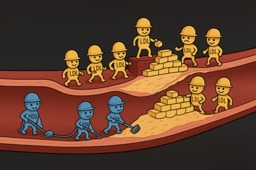
- LDL (Low-Density Lipoprotein): The “builder,” transporting cholesterol from liver to tissues for membrane construction and hormone production. Its moderate size allows it to penetrate arterial walls — where it becomes dangerous if oxidized, triggering plaque formation[4].
Painted by AI
- HDL (High-Density Lipoprotein): The “janitor,” collecting excess cholesterol and bringing it back to the liver for disposal. It also penetrates arterial walls — but helps clean them up, rather than gunk them up[5].
So LDL-C refers specifically to cholesterol carried by LDL particles. The “-C” matters. LDL (no “-C”) refers to the carrier. Same goes for HDL and HDL-C.
Chylomicrons and VLDL are too large to penetrate vessel walls and don’t form plaque. HDL actually reduces plaque risk. Only LDL crosses into danger territory.
That’s why managing blood lipids means managing LDL-C.
Also: elevated fasting triglycerides (TG) matter too. Why? Because TG travels via VLDL, which later converts to LDL. High TG is a warning sign that your body is upstream in the LDL pipeline.
Bottom line: LDL-C is the critical marker for cardiovascular risk.
2. Plaque Formation: How and Where It Starts
Think of blood vessels like plumbing. If the “water” is dirty — i.e., filled with oxidized LDL — gunk builds up. This triggers inflammation and the gradual formation of atherosclerotic plaque. If the plaque ruptures or dislodges, it can cause clots that trigger heart attacks or strokes.
Key point: if plaque is found in the carotid artery (a shallow, easily scanned area), there’s a good chance it exists elsewhere too — like coronary, cerebral, or renal arteries. Carotid plaque is a visible clue, not an isolated case.
And here’s the kicker: once formed, plaque is nearly impossible to reverse. Even the best interventions can only slow it down, not erase it.
3. The Real Culprit Behind High LDL-C: Sugar, Not Fat
I used to think eating too much fat was the problem. The deeper I looked, the clearer it became: the real driver is excess free sugars — particularly glucose and fructose in their isolated, rapidly absorbable forms.
Free sugars refer to monosaccharides and disaccharides (like glucose, fructose, sucrose) that aren’t bound to fiber, fat, or protein. They’re found in added sugars (e.g., soda, pastries, syrups) and natural concentrates (honey, fruit juice).
Sugars embedded in whole foods — fruits, veggies, grains, dairy — are absorbed slowly and don’t count as “free.”
Mechanism 1: Glucose spikes insulin, which prompts the liver to convert it into fat (TG), packages it into VLDL, and sends it off — eventually becoming LDL-C.
Mechanism 2: Fructose is metabolized almost entirely in the liver, and even more readily converted to fat. It skips the blood sugar spike — but still cranks up LDL-C.
Mechanism 3: Excess sugar leads to visceral fat accumulation, which releases fatty acids back to the liver, further fueling VLDL and LDL-C.
Add chronic inflammation and insulin resistance to the mix — both fueled by high sugar and visceral fat — and the cycle becomes self-perpetuating. Inflammation oxidizes LDL; insulin resistance prolongs its circulation.

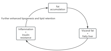
This feedback loop — sugar to fat to plaque — is how high LDL-C becomes a cardiovascular time bomb.
WHO recommends keeping free sugars below 5% of daily calories. That’s 25g on a 2000-calorie diet — less than one can of soda.

Painted by AI
In Summary: Hunting the True Killer
- Cardiovascular disease is the leading killer in China, driving ~50% of mortality.
- The culprit: atherosclerotic plaque.
- The enabler: high LDL-C.
- The upstream trigger: excess sugar → visceral fat → inflammation + insulin resistance.
- Triglycerides fuel LDL production via the VLDL pathway.
- Most actionable intervention? Cut sugar — especially refined, added sugars.
Call to Action: Slash Free Sugar Intake
If I could recommend just one intervention, it would be this: radically reduce your intake of free sugars — particularly fructose and sucrose.
This isn’t just an opinion; it’s backed by evidence[6–9]:
- Meta-analyses (Lancet): High sugar intake correlates with obesity, dyslipidemia, diabetes, and more. High fiber + low sugar = lower risk.
- U.S. guidelines: No added sugar for children under 2.
- Chinese dietary guidelines: Ideally keep added sugar <25g/day.
Addendum 1: LDL-C Is a Structural Builder — Could Lowering It Too Much Backfire?
Some sharp-eyed readers may wonder if our current approach to managing LDL-C is too rigid — perhaps even missing the forest for the trees. After all, LDL is the body’s main cholesterol courier, and cholesterol itself is essential for building cell membranes, synthesizing hormones, and producing vitamin D. It’s a foundational material for life.
So clearly, LDL-C isn’t just a villain. The real question is:
- If the body has high repair demands, could driving LDL-C too low be risky?
- Or more fundamentally — could elevated LDL-C actually reflect increased repair needs rather than just poor metabolic health?
- Should we be managing the number — or addressing the underlying causes of damage that drive the need for cholesterol?
To unpack this, we need to look at LDL-C regulation through two lenses: demand and supply.
On the demand side, LDL delivers cholesterol to tissues for cell membrane repair and renewal. LDL doesn’t “seek out” damaged tissue, but when tissues are healing, they upregulate LDL receptors — effectively pulling in more cholesterol for repair. That’s a demand-driven uptake.
And what drives damage? Chronic inflammation and oxidative stress — both of which are tightly linked to high-sugar diets. So in some cases, sugar doesn’t just create LDL — it also creates the need for LDL.
But here’s the twist: despite LDL-C’s transport role, the body’s actual cholesterol requirements are quite modest. Most cells can synthesize their own. So unless someone is severely deprived, having lower LDL-C rarely results in functional deficits.
Multiple studies have shown that even when LDL-C is lowered to below 0.78 mmol/L (30 mg/dL), there’s no evidence of cognitive decline, bleeding risk, or increased infection in the short to medium term[10–14]. While ultra-long-term data (spanning decades) is still being gathered, current clinical guidelines (e.g., <1.8 mmol/L) are considered safe and strongly beneficial.
Now the supply side: High-sugar diets stimulate the liver to produce triglycerides, which are packaged into VLDL particles for export. As VLDL circulates, it sheds triglycerides and morphs into LDL. The more VLDL your liver pumps out, the more LDL ends up in circulation. In this sense, high LDL-C is a supply-side issue driven by diet — especially sugar, not fat.
Which brings us full circle: Sugar doesn’t just increase the body’s need for LDL (via inflammation and repair). It also cranks up the production of LDL (via liver fat synthesis).
That dual mechanism makes sugar a metabolic double agent.
And it clarifies something crucial: rising LDL-C isn’t necessarily a useful or “adaptive” healing response. More often, it reflects systemic dysfunction: excess substrate, chronic inflammation, disrupted feedback loops.
Addendum 2: Why Blame Sugar — Not Fat — for Elevated Triglycerides?
When we check lipid panels during physicals, we’re often asked to fast beforehand. Why? Because we’re measuring fasting triglycerides — a specific metric that reflects what’s happening in the body after a night of metabolic processing.
Indeed, it’s a metabolic crystal ball that hints at your LDL-C trajectory.
Sure, triglycerides spike after meals — especially if you’ve eaten fatty foods. But that rise is short-lived, driven by chylomicrons (large particles that ferry dietary fat into the bloodstream). Within hours, tissues absorb that fat, and levels normalize.
Fasting triglycerides are different. After a night of digestion, these levels tell us what your liver’s been up to — specifically, whether it’s converting excess sugars into fat and packaging it into VLDL. That’s what we call endogenous fat production — and it’s a signature of metabolic trouble.
So when fasting TG is high, it’s not because you had bacon last night. It’s because your liver has been moonlighting as a fat factory. And that’s the precursor to a flood of LDL-C.
In other words, high fasting TG signals internal fat manufacturing. A yellow flag on your metabolic dashboard.
The danger isn’t how much fat you eat. It’s whether your liver has been forced into fat production mode — which is exactly what sugar excels at provoking.
Disclaimer: This article does not constitute medical advice. It reflects personal studies, researches, practices and experiences, and is for reference only. For medical guidance, please consult a physician or qualified healthcare professional.
Intervention recommendations are geared toward generally healthy individuals. If you have existing health conditions, always follow your doctor’s instructions.
References:
[1]Moschandrea, Chrysanthi, et al. “Mitochondrial Dysfunction Abrogates Dietary Lipid Processing in Enterocytes.” Nature, vol. 625, 2024, pp. 385–392.
[2]Dash, Satya, et al. “New Insights into the Regulation of Chylomicron Production.” Annual Review of Nutrition, vol. 35, 2015, pp. 265–294.
[3]Duan, Yajun, et al. “Regulation of Cholesterol Homeostasis in Health and Diseases: From Mechanisms to Targeted Therapeutics.” Signal Transduction and Targeted Therapy, vol. 7, 2022, article 265.
[4]Libby, Peter, et al. “Atherosclerosis.” Nature Reviews Disease Primers, vol. 5, 2019, article no. 56.
[5]Pownall, Henry J., et al. “High-Density Lipoproteins, Reverse Cholesterol Transport and Atherogenesis.” Nature Reviews Cardiology, vol. 18, no. 10, 2021, pp. 712–723.
[6]World Health Organization. Guideline: Sugars Intake for Adults and Children. WHO, 2015.
[7]Reynolds, Andrew, et al. “Carbohydrate Quality and Human Health: A Series of Systematic Reviews and Meta-analyses.” The Lancet, vol. 393, no. 10170, 2019, pp. 434–445.
[8]U.S. Dept. of Health and Human Services and U.S. Dept. of Agriculture. Dietary Guidelines for Americans, 2020–2025. 9th ed., Dec. 2020.。
[9]Chinese Nutrition Society. Dietary Guidelines for Chinese Residents (2022). People’s Medical Publishing House, 2022.
[10]Huynh, Karina. “PCSK9 Inhibition Reduces Cardiovascular Events in High-Risk Patients.” Nature Reviews Cardiology, vol. 14, 2017, p. 251.
[11]Fernández-Ruiz, Irene. “No Effect of PCSK9 Inhibitors on Cognitive Function.” Nature Reviews Cardiology, vol. 14, 2017, p. 568.
[12]Lim, Gregory B. “No Limit to the Benefits of LDL-Cholesterol Lowering.” Nature Reviews Cardiology, vol. 12, 2015, p. 444.
[13]Bao, Xuhui, et al. “Targeting Proprotein Convertase Subtilisin/Kexin Type 9 (PCSK9): From Bench to Bedside.” Signal Transduction and Targeted Therapy, vol. 9, art. 13, 2024.
[14]Emdin, Connor A., et al. “Analysis of Predicted Loss-of-Function Variants in UK Biobank Identifies Variants Protective for Disease.” Nature Communications, vol. 9, art. 1613, 2018.
메타데이터
- post_id
- fb63f7ac1bbb
- slug
- start-with-the-killer-that-takes-half-of-us-why-blood-lipids-are-my-entry-point-for-intervention-fb63f7ac1bbb
- url
- https://medium.com/@primeforever/start-with-the-killer-that-takes-half-of-us-why-blood-lipids-are-my-entry-point-for-intervention-fb63f7ac1bbb
- canonical_url
- https://medium.com/@primeforever/start-with-the-killer-that-takes-half-of-us-why-blood-lipids-are-my-entry-point-for-intervention-fb63f7ac1bbb
- author_url
- https://medium.com/@primeforever
- status
- ok
- fetched_at
- 2026-06-14 13:58:26