Fact-Checking: Bioaccumulation of microplastics in decedent human brains (Nature Medicine, 2025)
https://www.nature.com/articles/s41591-024-03453-1
Fact-Checking: Bioaccumulation of microplastics in decedent human brains (Nature Medicine, 2025)
https://www.nature.com/articles/s41591-024-03453-1
Validity of Microplastic Detection Methods
The detection techniques used in the study – Pyrolysis Gas Chromatography – Mass Spectrometry (Py‐GC/MS), Fourier-transform infrared spectroscopy (FTIR), and electron microscopy with energy-dispersive X-ray spectroscopy (EDS) – are generally accepted tools for identifying micro- and nanoplastics, including in biological tissues. Py‐GC/MS is a well-established analytical method that heats samples to break polymers into characteristic fragments, allowing identification and quantification of plastic polymers by their unique mass spectra. In fact, Py-GC/MS has shown good inter-laboratory consistency in detecting microplastics from human tissues and has been successfully applied in prior studies of human organs (e.g. placenta and arteries) . The Nature Medicine authors themselves note that their Py-GC/MS approach, while new to brain tissue, yielded comparable data across two independent labs, instilling confidence in its reliability . They also cite prior validations of Py-GC/MS for human samples, reinforcing it as a credible technique for microplastic analysis .
FTIR spectroscopy (including micro-FTIR and ATR-FTIR) is another widely used method for microplastic identification. It detects the unique vibrational signatures of polymer bonds, confirming plastic types. In the brain study, a subset of samples was analyzed by ATR-FTIR, which confirmed that polyethylene (PE) was the predominant polymer present . This use of FTIR is consistent with other research – for example, a 2024 JAMA Network Open case series examined human olfactory bulb tissues with micro-FTIR and likewise detected microplastics in 8 of 15 individuals . The JAMA study identified particles and fibers (mostly polypropylene) via their infrared spectra , demonstrating that FTIR-based techniques are indeed viable for finding and identifying microplastics in human brain tissue.
The study also employed electron microscopy (SEM and TEM) for visualizing the particles, coupled with EDS to analyze elemental composition. Electron microscopy is a standard tool to observe the morphology and size of micro/nanoparticles. In this case, SEM imaging revealed tiny shard-like particles in the tissue, and SEM-EDS confirmed these particles were composed primarily of carbon – consistent with plastic material (since inorganic contaminants would show metal or mineral elements). Transmission EM further visualized nanoscale fragments (<200 nm) from brain tissue digests , indicating the presence of nanoplastics that are too small for light microscopy. While EDS cannot conclusively identify polymer type (it only shows elements like carbon), it bolsters the evidence that the observed particles are organic. Combining these methods provides complementary strengths: Py-GC/MS and FTIR give chemical identification of polymer types, and microscopy gives physical confirmation of particles in the tissue. External experts have noted that Nihart et al. used a “state-of-the-art and complementary” suite of methods (Py-GC/MS, SEM-EDS, ATR-FTIR) which strengthens the credibility of the findings by cross-verifying the presence of microplastics through multiple approaches . In summary, the detection techniques are widely accepted in microplastics research and, when carefully executed with contamination controls, are considered reliable for identifying micro- and nanoplastics in human tissues.
Core Findings in Context of Other Studies
Key findings of the Nature Medicine study include: (a) microplastics were found in human brain tissue (frontal cortex) at measurable concentrations, with polyethylene (PE) being the most abundant polymer; (b) microplastic loads in brain (and liver) samples from 2024 were significantly higher than those in samples from 2016; and (c) brains of individuals with dementia had markedly higher microplastic levels than those of individuals without dementia. We evaluate each of these findings against the broader scientific literature and other peer-reviewed studies:
• Microplastics in human brain tissue: This study was among the first to quantitatively document microplastic accumulation in the human brain. Its discovery is supported by emerging evidence from other research. Notably, a 2024 study in JAMA Network Open analyzed the olfactory bulbs of human cadavers and found microplastics in more than half of the cases . The predominant polymers in that case were polypropylene fibers and particles , suggesting that inhaled microplastics can travel via the olfactory nerve pathway into the brain. This complements the Nature Medicine finding of plastics in the frontal cortex, indicating that multiple entry routes (ingested or inhaled) might deposit microplastics in different brain regions. Additionally, microplastics have been detected in other human tissues with direct environmental interfaces – for example, in lung tissue and in placentas – reinforcing the plausibility that some of these particles migrate systemically. A landmark 2021 study by Ragusa et al. found microscopic plastic fragments in human placentas , and plastics were even detected circulating in human blood in a 2022 study . Finding micro- and nanoplastic particles in the brain is consistent with these reports of plastics permeating the human body. While prior to 2023 there was little direct evidence of brain accumulation in humans, animal studies had already shown that micro- and nanoplastics can cross physiological barriers. Experiments in rodents have demonstrated that ingested nanoplastics can enter the bloodstream and even cross the blood – brain barrier, depositing in brain tissue and triggering inflammation . For example, polystyrene microplastics fed to mice were recovered in the brain within hours and were associated with neuroinflammatory responses and behavioral changes . Such studies substantiate the biological feasibility of the Nature Medicine finding. In short, the detection of microplastics in human brain tissue is credible and in line with a growing body of evidence that these particles are present throughout the human body and can reach even well-protected organs like the brain.
• Predominance of polyethylene (PE) in brain microplastics: Nihart et al. reported that ~75% of the plastic mass in brain samples was polyethylene, a common plastic polymer . They noted that PE was disproportionately accumulated in brain compared to other polymers, unlike in liver or kidney which showed a more even mix . This result is plausible given environmental exposure patterns – PE is one of the most abundantly produced plastics (used in bags, packaging, etc.) and is a major component of plastic pollution worldwide. Expert commentary has pointed out that PE is indeed “the most widely encountered polymer in environmental plastic litter” and is heavily used in food packaging, so its high abundance in human tissues “reflects its abundance in wildlife samples” as well . In other words, the predominance of PE in the brain mirrors what is found in the environment and food chain. Other human studies have found different polymers depending on the exposure route and tissue examined. For instance, the olfactory bulb study found mostly polypropylene , possibly due to inhalation of synthetic fibers (like those from textiles or masks) which are often PP. Microplastics in blood were reported to include polyethylene terephthalate (PET) and styrene polymers , which could reflect ingestion of bottled water or inhalation of polystyrene dust. Thus, while not every study finds PE as the top polymer, the high PE content in the brain is not implausible; it likely indicates that many of the plastic particles crossing into the brain derive from ubiquitous PE sources (perhaps tiny fragments of packaging materials in food or water). The Nature Medicine authors strengthened this claim by confirming PE chemically via FTIR . Overall, the polymer profile (dominated by PE, with smaller contributions of polypropylene, PVC, styrene-butadiene rubber, etc.) is consistent with common plastics in the environment , lending credibility to the idea that these brain microplastics originate from general environmental exposure.
• Higher concentrations in 2024 vs. 2016 samples: The study found a significant increase (roughly 50% higher on average) in total microplastic burden in brains collected in early 2024 compared to those from 2016 . This temporal trend is supported by external data on rising environmental microplastic pollution. As plastic production and use have climbed in the last decade, so too have human exposure levels. Professor Tamara Galloway, an expert in microplastic ecotoxicology, noted that the 50% increase over 8 years aligns with the steep increase in global plastic production and waste during a similar timeframe . Indeed, global plastic pollution has been growing exponentially, and microplastic contamination in water, air, and food has likely intensified between 2016 and 2024. Although no other human study has directly measured microplastics over time in stored tissue, the Nature Medicine team bolstered this finding by examining archived brain specimens from the late 1990s and early 2000s. Those older samples (1997 – 2013) showed significantly lower microplastic levels (median ~1,254 µg/g) than the more recent 2010s samples . Despite coming from different geographic locations, the older brains support the notion of rising microplastic exposure over the past decades . While ideally one would have continuous yearly data, the available evidence does not contradict the reported trend – if anything, it’s expected. No peer-reviewed study has challenged this specific temporal finding; given what we know about environmental pollution, most experts find it plausible that human microplastic burdens are increasing with time. This provides a strong incentive, as Galloway remarked, that reducing environmental plastic emissions could reduce human exposure in the future .
• Higher microplastic levels in dementia cases: Perhaps the most provocative observation was that brains from individuals with diagnosed dementia had significantly higher microplastic concentrations (median ~26,000 µg/g) – on the order of 5 – 10 times more than age-matched “normal” brains . This correlation is intriguing but must be interpreted cautiously. Importantly, the study does not claim that plastics cause dementia. The authors themselves emphasize that no causality can be concluded; rather, they speculate that dementia-related physiological changes (such as blood-brain barrier breakdown and impaired clearance mechanisms) could lead to greater accumulation of particles in the brain . This interpretation is consistent with known dementia pathology: conditions like Alzheimer’s disease often involve a leaky blood-brain barrier and reduced waste clearance (e.g. dysfunctional glymphatic system). External experts agree that the dementia association is likely due to such factors. As Dr. Matthew Campen (the study’s senior author) stated, patients with dementia may “simply accumulate more [microplastics] due to the disease process itself” rather than the plastics causing the disease . To date, there are no other human studies that directly compare microplastic loads in neurodegenerative disease vs. normal brains, so this finding stands on its own for now. It is neither directly supported nor refuted by prior literature, since it’s a novel observation. However, indirect support comes again from animal studies: in rodent experiments older animals were found to suffer more severe effects from microplastic exposure than younger ones , hinting that an aged or compromised brain might retain more particles or be more susceptible to their effects. Some environmental health researchers have hypothesized that chronic exposure to airborne particulates (including microplastics) could contribute to neuroinflammation, which is a risk factor for dementia. That remains unproven, but the concept is biologically feasible. The key point is that while dementia brains in this study did contain extraordinarily high microplastic levels, this result is viewed as a correlation. It raises an important question for further research – could long-term microplastic exposure exacerbate neurodegenerative disease? – but it does not by itself demonstrate a cause-and-effect relationship . Scientists urge further studies (and larger sample sizes) to see if this pattern holds and to investigate the mechanistic links.
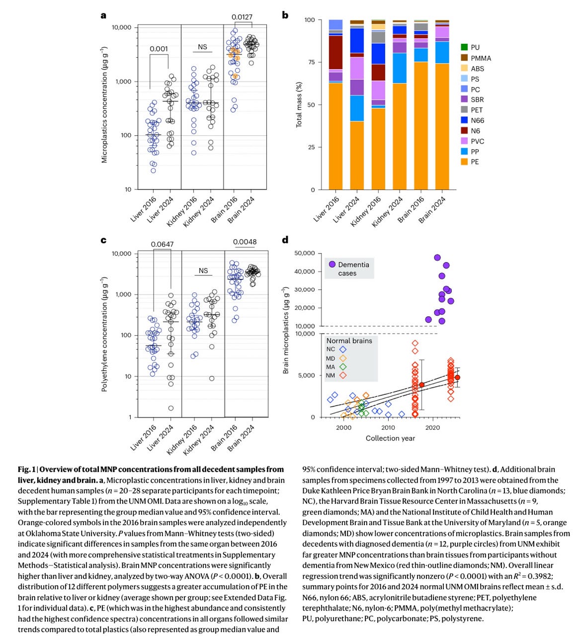
Figure 1: Key findings from Nihart et al. (2025) on microplastics in human organs . (a) Microplastic concentrations (µg of plastic per gram of tissue, log scale) in liver, kidney, and frontal cortex brain samples from 2016 (blue) and 2024 (black) autopsies. Brain tissues had much higher levels than liver or kidney in both years (note the brain’s median is ~3,300 µg/g in 2016 and ~4,900 µg/g in 2024, vs. a few hundred µg/g in liver/kidney) . Levels in 2024 were significantly greater than in 2016 for both brain and liver (p values shown) . (b) Composition of plastic by polymer type in each organ/year. Orange segments represent polyethylene (PE), which made up ~75% of the total plastic mass in 2016/2024 brains . In contrast, liver and kidney samples had more mixed polymers. (c) Concentrations of just PE in the organs, showing a similar pattern to total microplastics (brain ≫ other organs, and increasing over time) . (d) Brain microplastic levels over collection year. Colored diamonds are individual “normal” brains from various years and sources (1997 – 2013 from three brain banks, and 2016/2024 from New Mexico) . The black line indicates an upward trend over time. Purple circles (top right) are dementia cases (2019 – 2024) which cluster at much higher values than normal brains .
In summary, the core findings are generally in line with current scientific understanding. The presence of microplastics in human brains is supported by other studies and considered plausible by experts. The predominance of polyethylene is explained by its environmental ubiquity. The increasing trend from 2016 to 2024 is consistent with rising pollution levels, and experts find this credible . The extremely high levels in dementia patients, while not previously reported elsewhere, are cautiously interpreted and deemed plausible as an effect (rather than a cause) of neurodegeneration. No peer-reviewed research has yet outright contradicted these findings; given how recent this study is, it remains to be corroborated by follow-up investigations. But taken together with related studies, the evidence leans toward validating the study’s main conclusions (with appropriate caveats about causation).
Scientific Commentary and Peer Reactions
The publication of this study in Nature Medicine drew significant attention and a mix of reactions from the scientific community. Several independent experts have weighed in, offering perspectives that both affirm the importance of the findings and highlight uncertainties. Below is a summary of key commentary:
• Novelty and Significance: Researchers have largely acknowledged the study as an important first step in mapping microplastics in internal organs. Dr. Antonis Myridakis (Brunel University) commented that the work provides “compelling evidence” that micro- and nanoplastics can cross the blood – brain barrier and accumulate in human brain tissue, especially highlighting the finding of predominant polyethylene in the brain . He noted that the authors used robust, complementary methodologies, which strengthens confidence in the results . This sentiment – that the multidisciplinary approach is a technical advancement – was echoed by others. It addresses a known difficulty in the field: until now, detecting plastic particles in internal organs was challenging due to lack of established methods . By overcoming some of these analytical hurdles, the study is seen as a valuable contribution. Prof. Tamara Galloway (University of Exeter) also emphasized that it’s not surprising to find microplastics in our bodies given their ubiquity in air, water, and food, but this study usefully maps how they may distribute in specific organs . She pointed out that the work makes the issue “personal” by showing tangible amounts in human brain tissue, underscoring the need to understand health implications .
• Alignment with Prior Evidence: Experts placed the findings in context, generally agreeing that they fit with existing evidence. Prof. Galloway remarked that the observed 50% rise in brain microplastic levels over 8 years is significant and “reflects the increased production and use of plastics over a similar timeframe” . This indicates that if society reduces microplastic pollution, human body burdens might also decrease – a hopeful note she adds . Dr. Myridakis similarly stated the study “aligns with recent findings that MPs/NPs are present in blood and major organs,” and he found the discovery of plastic particles in cerebrovascular walls and immune cells particularly insightful regarding potential neuroinflammatory roles . In other words, the community sees the results as part of a logical continuum of microplastic research.
• Calls for Caution and Confirmation: Despite the general enthusiasm for the topic, scientists urge caution in interpretation. Prof. Oliver Jones (RMIT University), an analytical chemist, noted that “exceptional claims need exceptional evidence” and advised not to jump to alarming conclusions without careful scrutiny . He and others pointed out that this is an initial study with a relatively limited sample size (brains from 52 individuals across two time points) . While the data are intriguing, more data – especially from other regions and continuous years – would be needed to firmly establish trends or generalize the findings . Both Prof. Jones and Prof. Theodore Henry (Heriot-Watt University) stress the need for independent confirmation of the results . They suggest that other researchers should attempt to detect microplastics in archived human tissues to validate the presence and levels reported. Prof. Henry found it “surprising that similar particles have not been detected in other studies or examinations of the same tissues” until now, given the high concentrations reported . This doesn’t mean the results are wrong, but it underscores that replication is important. Encouragingly, the Nature Medicine team did include a partial independent replication – five brain samples were re-analyzed by a separate lab using the same Py-GC/MS method and yielded consistent values . This cross-check within the study itself adds credibility, but the broader community still looks forward to external groups repeating such analyses.
• No Overstatement of Health Effects: Commentators have also lauded the authors for not overstating the health implications. The study explicitly did not claim that microplastics cause neurological disease, and experts agree with this cautious stance. “As the authors themselves note, there is as yet no strong evidence of any health effects” from these brain microplastics at the observed levels, Prof. Jones emphasized . Dr. Myridakis likewise highlighted that no causality was established between brain plastic load and dementia or any other health outcome . The consensus is that it’s an important finding to know these particles are there, but any link to disease remains speculative. Further research will need to investigate whether chronic microplastic accumulation has subtle effects on brain tissue – for instance, contributing to inflammation – or if they are inert bystanders. Until such data are available, experts caution against alarmism.
In summary, the peer reaction has been measured: scientists find the study credible and valuable, but they also identify it as an early foray that needs follow-up. There is broad agreement that microplastics in organs is an issue of concern and worthy of study , but also an understanding that methods to detect them are complex and results must be confirmed and expanded. No one has fundamentally refuted the findings, but several have pointed out that alternate explanations (analytical artifacts or contamination) must be ruled out (discussed below) before fully accepting the conclusions. This balanced reaction is common for novel results – intrigue and cautious optimism, coupled with calls for further verification.
Methodological Limitations and Potential Sources of Error
While the study’s methods were cutting-edge, scientific critics have highlighted several limitations and possible error sources that could affect the accuracy of the results. It’s important to consider these when assessing the study’s credibility. The major points of concern include:
• Analytical Interference (False Positives): A key issue is that brain tissue is rich in fats (lipids), and these could interfere with Py-GC/MS measurements. Pyrolysis GC/MS works by breaking compounds into fragments – but certain lipids can degrade into hydrocarbon fragments that resemble those from polyethylene. Prof. Oliver Jones cautioned that “the brain is mainly made of fat” and fats can yield the same pyrolysis products as polyethylene, the main polymer reported . This raises the possibility that some fraction of what was measured as “PE” might actually be signals from residual biological material, not plastic. The authors did recognize this concern: in their discussion, they acknowledge that “lipids have been noted as a potential source of interference in Py-GC/MS analysis of PE” and that unknown residual biomolecules in the samples could cause overestimation. To mitigate this, they performed an extensive KOH digestion to dissolve organic matter; KOH eliminated >99% of organ mass in liver/kidney and ~92% in brain samples . The less complete digestion of brain (owing to its high fat content) means more non-plastic residue remained (about 8% of the original mass) . The team chose not to use organic solvents (which might extract lipids along with plastics) and instead relied on physical separation to remove fats . While these steps likely reduced many interferences, some uncertainty remains. The authors observed that their spectra for brain showed fewer long-chain hydrocarbons than expected for pure PE, possibly because brain lipids or oxidized plastics altered the signature . This complexity means there is a margin of error in the absolute quantities reported. External analysts suggest results should be confirmed with alternative methods (e.g. mass spectrometry of specific markers or direct imaging techniques) to ensure that what was measured as polymer mass is not an artifact of brain lipid pyrolysis. In short, false positives for PE due to fats are a known pitfall , and the study took reasonable steps to address it, but it remains a critical point for follow-up validation.
• Contamination Controls: Detecting microplastics at trace levels is notoriously prone to contamination – airborne fibers, synthetic dust, or plastic tools in the lab can all introduce spurious particles. The authors implemented numerous quality control measures to minimize contamination. They ran blank samples (e.g. KOH alone, and the formalin solution used to store tissues) through the entire process to check for any plastic signals – none were significant . They also catalogued the polymer composition of all labware (tubes, pipette tips) used, to distinguish those materials from sample results . All samples (2016 and 2024, all organs) were handled under consistent protocols in the same medical examiner environment, reducing variability in external contamination . Notwithstanding these precautions, experts note that contamination is difficult to 100% rule out. Prof. Jones argued that using consistent protocols doesn’t guarantee no contamination – it could just mean any contamination that did occur would affect all samples similarly . For example, if a certain step introduced plastic fibers from the air or from a piece of equipment, every sample might pick up similar contamination, potentially misleadingly “consistent” results . He gave a concrete example: disposable lab gloves can shed microplastic particles and cause false positives . (Indeed, a 2020 study showed that improper glove use led to erroneous microplastic findings in samples .) Another concern is the autopsy process itself – bodies are often stored and examined on plastic surfaces. Jones pointed out that body bags are made of polyethylene ; if brain tissues were in contact with such bags or plastic jars, that could theoretically introduce PE fragments. The authors counter this by noting that older samples (stored ~7 years in plastic) actually had lower plastic levels than newer samples stored briefly , which suggests storage containers didn’t leach significant plastic into the tissues. Nonetheless, ambient microplastics are “almost everywhere,” as Jones emphasized, and ensuring that detected particles truly originated in vivo rather than from the lab or autopsy room is a continual challenge . Future studies will need to replicate these findings possibly using clean-room conditions or non-plastic equipment (as some researchers recommend ) to absolutely confirm that the brain plastics are not an artifact. So far, there is no clear evidence of a contamination error in this study, but the possibility of consistent low-level contamination cannot be entirely dismissed.
• Sample Size and Representation: The study analyzed tissues from dozens of individuals (28 brains from 2016, 24 brains from 2024, plus 13 older brain-bank samples and 12 dementia cases). While this is quite substantial for a study of this kind, it still represents a limited slice of the population (mostly people from New Mexico, USA, in certain years). Prof. Jones noted that “there is not enough data to make firm conclusions on the occurrence of microplastics in [brains] of New Mexico, let alone globally” . The time-trend (2016 vs 2024) is based on just two snapshot years, which is not a continuous trend – intermediate years weren’t analyzed, so one must be careful in interpreting a linear increase . Additionally, the dementia comparison involved 12 cases with mixed types of neurodegenerative disease ; this small number means any statistical conclusions there are preliminary. The study did a good job controlling for basic demographics (the 2016 vs 2024 groups had similar age ranges and causes of death, and no age correlation was found with plastic load ). However, as Dr. Myridakis pointed out, lifestyle factors were not accounted for . Individuals’ occupations, diets (e.g. high seafood consumption could elevate microplastic intake), living environment (urban vs rural air quality), etc., were unknown and could influence their microplastic exposure . Such factors might confound the differences observed. For example, if the 2024 cohort happened to include more people from high-pollution areas than the 2016 cohort, that might partly explain higher levels. The authors couldn’t control for all these variables with the available data. Thus, while the findings are statistically significant, caution is warranted in generalizing the exact values to all populations. More studies with larger and more diverse samples would firm up these results.
• Lack of Particle Characterization: The use of Py-GC/MS provides polymer mass but no information on the size, number, or shape of particles in each tissue. Prof. Henry highlighted this limitation: by its nature, pyrolysis “disintegrates” the polymers, so one cannot tell whether the plastic was a few large fragments or billions of nanoparticles . Particle size is crucial for understanding uptake mechanisms and potential toxicity. The authors did attempt to characterize particles via microscopy on a subset of samples, finding mostly <1 µm fragments in brain . But this was qualitative. The study essentially quantifies total polymer mass per gram of tissue – around 5 mg of plastic per gram of brain in recent samples – which is quite high (0.5% by mass). It remains to be confirmed how those grams of plastic are distributed (as countless nano-sized specks, or as some larger pieces). If mostly nanoscale, as the study suggests, some could have been lost during sample prep (the authors acknowledge ultracentrifugation might not pellet the tiniest particles, potentially leading to underestimation of total plastic mass ). On the other hand, if any large particles (>100 µm) were present, Py-GC/MS would detect them but the microscopy survey might have missed them due to sampling a small tissue area. The inability to fully visualize and count the particles leaves a gap in corroborating the mass measurements. Going forward, combining pyrolysis results with techniques like micro-FTIR mapping or Raman microscopy on tissue sections could help verify that the polymer masses correspond to actual discrete microplastics in the brain and not diffuse polymer contaminants or dissolved molecules.
• Absence of “blank” human tissues: One practical challenge is that in today’s world it may be impossible to find a truly microplastic-free control tissue. Every organ sample, including those from 1990s, contained some level of plastic in this study. Prof. Henry noted that essentially all tissues had plastic polymers detected, which means the researchers couldn’t compare exposed vs unexposed individuals . This ubiquity raises the stakes for ruling out lab contamination – if even older specimens have plastics, it suggests widespread exposure, but it also means any baseline “noise” in the method will appear as a positive result in every sample. Henry warned that this makes it difficult to be sure some low-level readings aren’t due to artifacts . It also complicates questions about health effects, since we lack an unexposed control group of people with zero plastic to see if having plastics correlates with any pathology. The omnipresence of plastics in modern life is both the motivation for the study and a hurdle for experimental design.
In conclusion, the study is scientifically credible but not without limitations. External experts have identified plausible sources of error – especially the possibility of analytical interference and contamination – that could influence the results. The researchers took many appropriate precautions (chemical digestion, blanks, cross-lab validation) to ensure accuracy , and they openly discussed the limitations in their paper . The consensus is that the findings are likely real but still preliminary. Future investigations should aim to replicate the results with even more stringent contamination control (perhaps in a clean-room setting), alternative analytical techniques (to confirm polymer ID and measure particle sizes), and larger sample sets. By addressing these limitations, the scientific community can more definitively validate how much plastic is truly accumulating in our brains and what, if anything, it means for human health .
Conclusion
Overall, the Nature Medicine article presents a carefully conducted study using advanced methods to detect micro- and nanoplastics in human organs, with a focus on the brain. Its detection methods (Py-GC/MS, FTIR, electron microscopy) are well-founded and generally reliable for this purpose, although like any technique they require rigor to avoid false positives. The core findings – that human brains contain measurable microplastics (especially PE), that levels have risen in recent years, and that dementia brains showed higher burdens – are credible and in line with broader evidence, though they need to be confirmed by further research. Other peer-reviewed studies (in human olfactory bulbs and in blood, placenta, etc., as well as animal models) support the notion that microplastics can enter the body and even reach the brain . Experts have reacted positively to the significance of the work but urge caution: the data, while important, are initial and should not be over-interpreted in terms of health impact without more evidence . Valid critiques have been raised regarding potential methodological shortcomings like lipid interference and contamination, which the authors partly addressed but not entirely eliminate. These limitations underscore that this finding – microplastics in the brain – though alarming, must be followed by replication and deeper investigation.
In summary, the study by Nihart et al. appears scientifically sound in its approach and its results are taken seriously by the scientific community, but it is not the final word on the topic. It serves as a pioneering report that opens up many questions: How widespread is plastic accumulation in human brains? What sizes and types of particles are most common? Could these particles contribute to diseases like dementia, or do they simply accumulate without effect? The article has high credibility as a peer-reviewed, methodically robust piece of research, and its conclusions are cautiously regarded as plausible. However, scientists agree that further work – with refined methods and consideration of the noted limitations – is essential to fully validate and understand the implications of finding microplastics in our brains . The study has effectively sounded an alarm and set the stage for ongoing research into the human health dimensions of microplastic pollution.
메타데이터
- post_id
- 1650e349dc10
- slug
- fact-checking-bioaccumulation-of-microplastics-in-decedent-human-brains-nature-medicine-2025-1650e349dc10
- url
- https://medium.com/@axegggl/fact-checking-bioaccumulation-of-microplastics-in-decedent-human-brains-nature-medicine-2025-1650e349dc10
- canonical_url
- https://medium.com/@axegggl/fact-checking-bioaccumulation-of-microplastics-in-decedent-human-brains-nature-medicine-2025-1650e349dc10
- author_url
- https://medium.com/@axegggl
- status
- ok
- fetched_at
- 2026-07-20 09:01:03