The Ghost Pixels: How AI Predicts Diseases from Scans, the Dark Bias of Medical Data, and the Human…
Introduction: When a Computer Develops a Medical Eye
The Ghost Pixels: How AI Predicts Diseases from Scans, the Dark Bias of Medical Data, and the Human Gatekeepers
Introduction: When a Computer Develops a Medical Eye

Imagine you are an expert radiologist with decades of experience. You sit in a dimly lit room, staring intensely at a patient’s chest X-ray. To your highly trained eyes, the lung tissue looks completely normal, clear, and healthy. You are about to sign off on a clean bill of health.
But then, you run the image through an Advanced Artificial Intelligence (AI) screening software. Within three seconds, the screen flashes. The AI highlights a microscopic, faint cluster of pixels in red, attaching a terrifying prognosis: “High probability of early-stage malignancy. Tumor development predicted within 6 months.”
How can a machine see what human eyes, backed by decades of medical school, completely miss? What is happening beneath the hood of this technology? More importantly, what are the hidden dangers of importing Western-trained AI models into Asian healthcare systems? Let’s pull back the curtain on the silent revolution taking over radiology.
1. The Anatomy of a “Ghost Pixel”: How AI Deep-Learns Imaging
To a human doctor, an X-ray or an MRI is a visual map of anatomy — bones, organs, and shadows. To an AI, that same image is nothing more than a giant mathematical matrix of numbers, contrasts, and pixels.
- Computer Vision and CNNs: AI analyzes medical scans using a specialized architecture called Convolutional Neural Networks (CNNs). These networks mimic the human brain’s visual cortex, breaking an image down into layers, from raw edges and shapes to micro-textures.
- The Contrast Blindspot: When a cancer tumor or a neurological anomaly begins to form, its early footprint is practically invisible. It might manifest as a variance of just 1 or 2 pixels, shifting slightly from a dark gray to a marginally lighter gray. Because human biological vision has a natural contrast threshold, our brains structurally filter out these micro-variations.
- The Matrix Advantage: The AI doesn’t look at the picture holistically; it calculates the exact numerical density of every single pixel relative to its neighbors. By recognizing mathematical anomalies in pixel distribution, it spots the “Ghost Pixels” long before they cluster into a visible mass.
2. Time-Traveling Diagnostics: Predicting Diseases Before They Exist
Traditional radiology is reactive — you take a scan to find a disease that is already causing symptoms. AI is turning radiology into a predictive science.
- The 5-Year Heart Attack Forecast: Through a process called opportunistic screening, an AI can analyze a standard chest X-ray — originally taken just to check a patient’s lungs — and accurately calculate their risk of a cardiovascular event (Heart Attack) within the next five years. It achieves this by tracking micro-calcifications and subtle geometric narrowing in the walls of the blood vessels surrounding the heart, patterns too minute for standard human tracking.
- Pre-Symptomatic Alzheimer’s Tracking: In brain MRI scans, AI can measure structural gray-matter volume down to the millimeter. By comparing a patient’s brain topography against millions of historic datasets, it can identify microscopic tissue shrinkage (atrophy) years before the patient experiences their first bout of memory loss.
3. The Dark Truth: The West-to-East Bias Trap (The Demographic Nightmare)
This is the hidden crisis currently keeping medical researchers up at night. While Asian countries (like India and China) boast the largest populations on earth, the vast majority of cutting-edge medical AI models are trained on historical data from hospitals in the United States and Europe.
- The Training Bias: If an AI model is trained on 100,000 scans of predominantly Caucasian patients, it memorizes the baseline biology of that specific demographic.
- The Physiological Disconnect: Asian populations have fundamentally different biological baselines. For example, East and South Asian populations naturally exhibit variations in average bone mineral density (BMD)and heart-to-thorax ratios compared to Western populations.
- The Danger of False Realities: When a Western-trained AI looks at an Asian patient’s X-ray, it applies its Western baseline. It might look at a perfectly healthy Asian bone structure, misinterpret the natural density difference as a disease, and trigger a false positive for severe osteoporosis. This leads to Misdiagnosis, unnecessary medications, and massive emotional distress.
The Shortcut: How Western AI Got to Asia So Fast
If training an AI for a specific population takes years of local data collection, how are Asian hospitals using this tech right now? The answer is a machine learning shortcut called Transfer Learning.
Instead of building a new AI from scratch, companies take a model that already knows the basics of human anatomy (learned from Western data). They bring it to Asia and subject it to Fine-Tuning — feeding it a much smaller local batch of 5,000 to 10,000 Asian scans. This fast-tracks the software into the market, but it doesn’t entirely erase the underlying demographic bias, leaving a lingering margins of error.
4. The Human Gatekeepers: How Doctors Fact-Check Invisible Data
This brings us to a massive logical paradox: If AI is catching micro-pixel anomalies that are structurally invisible to the human eye, how can an Asian doctor double-check the machine? How can a human validate something they literally cannot see?
The medical world solves this using a strict, multi-tiered Collaboration Framework:
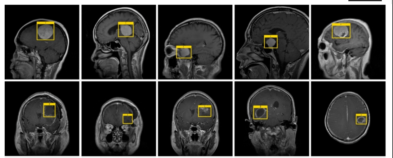
- The Red Flag System (Bounding Boxes & Heatmaps): The AI never just gives a text report saying “Cancer found.” It generates a visual overlay on the scan. It creates a bright Bounding Box or a glowing Heatmapdirectly over the exact coordinates of the suspicious pixels. It essentially tells the doctor: “Do not look at the whole lung. Zoom into these exact 4 millimeters.”
- Digital Weaponry: Once the doctor’s attention is locked onto that exact micro-region, they don’t rely on their naked eye. They use specialized diagnostic monitors and software to drastically alter the image’s contrast matrices, artificially amplifying the brightness and magnification of that specific zone to verify the anomaly.
- Clinical Correlation over Pixels: A computer only sees an image. A doctor sees a patient. The human physician correlates the AI’s red flag with the patient’s real-world clinical history: Is there a chronic cough? Genetic predispositions? Environmental exposures?
- Escalation Protocols: If the AI flags a pixel cluster that remains invisible on the X-ray even after zooming, a human doctor does not ignore it. Instead of risking a mistake, they use the AI’s flag as justification to order a higher-tier diagnostic test, such as a High-Resolution CT Scan, a PET Scan, or a Biopsy. The AI acts as the early warning radar; the human deploys the final confirmation tools.
5. The Black Box Enigma: The Ultimate Technical Roadblock
Even if the AI-doctor partnership works, healthcare faces one final, terrifying psychological wall: The Black Box Problem.
Deep learning neural networks process data through millions of interconnected mathematical nodes. The machine can tell you the final output (“This is a tumor”), but because the math is so incredibly complex and non-linear, it cannot explain its own reasoning. It cannot show the step-by-step logic of how it arrived at that conclusion.
If an AI makes a wrong prediction that leads to a patient’s injury, engineers cannot easily look inside the network to see exactly why it failed. Because of this liability, a multi-million dollar global race is underway to develop XAI (Explainable AI) — medical models engineered to not only find the disease but print out a human-readable roadmap explaining their medical rationale to the attending physician.
The Final Verdict: The Hound and the Hunter
Ultimately, Artificial Intelligence is not going to replace human doctors. Instead, it reframes the dynamic of modern medicine.
Think of AI as a highly trained bloodhound. It runs ahead into the brush, using its inhuman senses to sniff out microscopic, invisible threats hidden deep within the data matrix, barking loudly (marking the scan) when it finds a target. The human doctor is the hunter — the one with the wisdom, contextual empathy, and ultimate authority to decide whether to pull the trigger on a medical diagnosis.
By understanding the boundaries of its pixels, and fighting the biases in its data, humanity is entering an era where diseases are caught not when they harm us, but when they are just a whisper on a digital screen.
메타데이터
- post_id
- 378f133650cc
- slug
- the-ghost-pixels-how-ai-predicts-diseases-from-scans-the-dark-bias-of-medical-data-and-the-human-378f133650cc
- url
- https://medium.com/@gideonsivak2007/the-ghost-pixels-how-ai-predicts-diseases-from-scans-the-dark-bias-of-medical-data-and-the-human-378f133650cc
- canonical_url
- https://medium.com/@gideonsivak2007/the-ghost-pixels-how-ai-predicts-diseases-from-scans-the-dark-bias-of-medical-data-and-the-human-378f133650cc
- author_url
- https://medium.com/@gideonsivak2007
- status
- ok
- fetched_at
- 2026-07-27 23:02:56