From Transcriptomics to Protein Structure: Exploring AKT1 in Triple-Negative Breast Cancer Using…
Triple-Negative Breast Cancer (TNBC) remains one of the most aggressive and biologically complex subtypes of breast cancer. Unlike other…
From Transcriptomics to Protein Structure: Exploring AKT1 in Triple-Negative Breast Cancer Using AlphaFold2
Triple-Negative Breast Cancer (TNBC) remains one of the most aggressive and biologically complex subtypes of breast cancer. Unlike other breast cancer types, TNBC lacks expression of estrogen receptor (ER), progesterone receptor (PR), and HER2 amplification, which makes many targeted therapies ineffective. Because of this, researchers increasingly rely on transcriptomics, systems biology, and computational approaches to identify new molecular mechanisms and therapeutic opportunities.
In my recent bioinformatics project, I wanted to move beyond classical RNA-seq analysis and investigate how transcriptomic findings could be connected to structural biology. Instead of stopping at differential gene expression and hub gene identification, I explored what one of the most important TNBC-associated proteins might actually look like at the structural level.
This project became a bridge between transcriptomics, protein structure prediction, and structural bioinformatics.
The complete project, scripts, and visualisations are available on GitHub: 🔗 AlphaFold-TNBC GitHub Repository
Why AKT1?
Among the differentially expressed hub genes identified during my TNBC transcriptomics analysis, AKT1 attracted my attention because of its central role in the PI3K-Akt signalling pathway, one of the most dysregulated signalling networks in cancer biology.
AKT1 encodes a serine/threonine kinase responsible for regulating:
- Cell survival
- Cell proliferation
- Glucose metabolism
- Apoptosis suppression
- Growth signalling
- Protein synthesis
In many cancers, including TNBC, hyperactivation of the PI3K-Akt pathway promotes uncontrolled cellular growth and resistance to apoptosis. Because of this, AKT1 has become a major target in precision oncology and drug development research.
However, understanding gene expression alone is often insufficient. A gene may be highly expressed, but its biological behaviour depends heavily on the structure and flexibility of the encoded protein.
This is where structural bioinformatics becomes important.
Moving Beyond RNA-seq
Most transcriptomics projects typically end after identifying:
- Differentially expressed genes (DEGs)
- Functional enrichment pathways
- Protein-protein interaction networks
- Hub genes
But I wanted to continue one step further:
What structural properties allow AKT1 to behave as a highly dynamic oncogenic signalling protein?
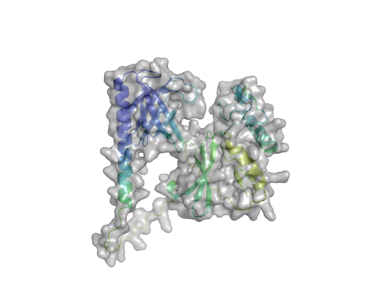
To investigate this question, I used AlphaFold2, one of the most transformative AI systems developed for protein structure prediction.
What is AlphaFold2?
DeepMind developed AlphaFold2, an artificial intelligence system capable of predicting protein 3D structures with unprecedented accuracy.
Protein structure prediction has historically been one of the biggest challenges in molecular biology because experimental techniques such as:
- X-ray crystallography
- Cryo-electron microscopy (Cryo-EM)
- Nuclear magnetic resonance (NMR)
are expensive, time-consuming, and technically demanding.
AlphaFold2 revolutionized the field by using deep learning architectures trained on massive structural datasets to infer atomic-level protein conformations directly from amino acid sequences.
For this project, I used:
- ColabFold v1.6.1
- MMseqs2-based sequence alignment
- AlphaFold2 prediction pipeline
Structural Prediction Workflow
The workflow included several major stages:
1. Protein Sequence Preparation
The amino acid sequence of AKT1 was prepared and submitted into the AlphaFold2 prediction pipeline using ColabFold.
2. Structure Prediction
AlphaFold2 generated predicted 3D conformations of AKT1 by combining:
- Evolutionary information
- Multiple sequence alignments (MSA)
- Attention-based neural networks
- Spatial geometry optimization
The final predicted structure included confidence metrics for every residue.
3. Confidence Analysis (pLDDT)
One of the most important AlphaFold metrics is:
Predicted Local Distance Difference Test (pLDDT)
0 \leq pLDDT \leq 100
This score estimates local structural confidence for each amino acid residue.
General interpretation:
- 90–100 → very high confidence
- 70–90 → reliable backbone prediction
- 50–70 → moderate confidence
- Below 50 → likely flexible or disordered regions
For AKT1:
\text{Mean pLDDT} = 65.61

This suggests that the model contains both confidently folded domains and flexible regions.
Interestingly, this is biologically meaningful rather than problematic.
Intrinsically Disordered Regions (IDRs)
One of the most interesting findings from the project was:
\text{IDR Content} = 30.2%
Intrinsically disordered regions (IDRs) are protein segments that do not adopt stable fixed conformations under physiological conditions.
Unlike rigid folded domains, IDRs remain flexible and dynamic.
For many years, these regions were mistakenly considered “unstructured noise.” Modern structural biology now recognizes that IDRs play critical regulatory roles in:
- Signal transduction
- Protein-protein interactions
- Molecular recognition
- Post-translational modification
- Allosteric regulation
In oncogenic proteins such as AKT1, structural flexibility can become biologically advantageous for cancer cells.
Flexible regions often act as:
- Interaction hubs
- Regulatory switches
- Dynamic binding surfaces

This may partially explain why AKT1 participates in numerous signalling pathways simultaneously.
Ramachandran and Secondary Structure Analysis
To better evaluate the predicted fold quality, I also performed secondary structure and Ramachandran analysis.
The results showed:
- 24.6% α-helices
- 32.9% β-strands
These findings are consistent with the classical bilobal kinase architecture observed in many signalling kinases.
The Ramachandran plot also supported acceptable stereochemical geometry across most structured regions.
This suggests that the predicted model is structurally plausible and biologically interpretable.
Contact Map Analysis
Another interesting part of the project involved contact map analysis.
A contact map visualizes residue-residue interactions within the protein structure.
The AKT1 contact map revealed:
- Distinct domain separation
- Long-range residue interactions
- Structural organization patterns
Importantly, three major domain blocks became visible:
- PH domain
- Kinase domain
- Regulatory domain
These domains correspond closely with experimentally characterized AKT1 architecture reported in the literature.
Connecting Multiple Layers of Biology
One of the most rewarding parts of this project was seeing how different biological layers could be integrated into a single workflow:
Transcriptomics → Network Biology → Structural Biology
The overall pipeline looked like this:
- RNA-seq differential expression analysis using DESeq2
- Hub gene identification through co-expression analysis
- Functional prioritization of AKT1
- Protein structure prediction using AlphaFold2
- Structural interpretation and disorder analysis
This integration helped transform transcriptomic data from abstract gene lists into biologically interpretable molecular systems.
Why Structural Bioinformatics Matters
Structural bioinformatics is becoming increasingly important in modern precision medicine because proteins are ultimately the functional molecules responsible for cellular behaviour.
Genes provide instructions, but proteins execute biological activity.
Understanding protein structure allows researchers to investigate:
- Drug binding mechanisms
- Mutation effects
- Conformational flexibility
- Active site accessibility
- Allosteric regulation
- Therapeutic targeting opportunities
As AI-driven structural prediction tools continue to improve, computational biology is increasingly capable of performing analyses that previously required expensive laboratory infrastructure.
Open Science and Reproducibility
One important goal of this project was maintaining transparency and reproducibility.
The entire workflow, including:
- Python scripts
- Visualisation notebooks
- Structural analysis outputs
- Prediction files
has been shared openly on GitHub.
Open science allows students, researchers, and independent learners to explore, reproduce, and improve computational biology workflows collaboratively.
GitHub Repository: 🔗 AlphaFold-TNBC GitHub Repository
Final Thoughts
This project further strengthened my interest in:
- Structural bioinformatics
- Cancer systems biology
- AI-driven molecular biology
- Computational oncology
It also showed me how modern bioinformatics is no longer limited to sequence analysis alone. Today, we can connect transcriptomics, network biology, machine learning, and structural prediction into unified biological interpretations.
I am especially inspired by scientists such as Garib Murshudov and Aynura Pashayeva, whose work motivated my growing interest in structural biology and computational research.
The future of cancer research will increasingly depend on interdisciplinary integration between AI, molecular biology, and bioinformatics — and projects like this demonstrate how accessible these technologies are becoming for independent researchers and students worldwide.
Bioinformatics #StructuralBiology #AlphaFold2 #CancerResearch #TNBC #ComputationalBiology #Transcriptomics #AI #OpenScience #GitHub
메타데이터
- post_id
- db6a5a3b17ce
- slug
- from-transcriptomics-to-protein-structure-exploring-akt1-in-triple-negative-breast-cancer-using-db6a5a3b17ce
- url
- https://medium.com/@suleyman.hacizade1/from-transcriptomics-to-protein-structure-exploring-akt1-in-triple-negative-breast-cancer-using-db6a5a3b17ce
- canonical_url
- https://medium.com/@suleyman.hacizade1/from-transcriptomics-to-protein-structure-exploring-akt1-in-triple-negative-breast-cancer-using-db6a5a3b17ce
- author_url
- https://medium.com/@suleyman.hacizade1
- status
- ok
- fetched_at
- 2026-06-09 15:37:30