Bioengineering Stem Cells for Creating Vital Organs Using Growth Factors
Thousands of people die each year waiting for a kidney that never comes. What if instead of waiting for a donor, doctors could grow one for…
Bioengineering Stem Cells for Creating Vital Organs Using Growth Factors
Thousands of people die each year waiting for a kidney that never comes. What if instead of waiting for a donor, doctors could grow one for you?
Introduction
Every year, more people are added to organ transplant waiting lists than there are organs available to give them. The kidney is currently the organ in highest demand. Right now, the only option is to wait for a compatible donor — and usually the wait is 3–5 years long. Regenerative medicine, a field focused on engineering or regenerating human tissues and organs, offers a different path.
The question this project investigates is: what growth factors can guide stem cells to develop into kidney cells? Answering that question is a necessary step toward building a patient-specific kidney outside the body and being able to transplant it — an organ with no donor required, a reduced risk of immune rejection, and a much shorter wait time.
Background
To understand the approach, a few terms are worth defining.
Stem cells are cells that have not yet committed to a specific identity. They can differentiate into virtually any cell type in the body. Human induced pluripotent stem cells (iPSCs) are adult cells that have been reprogrammed back into this undifferentiated state, making them a practical and straightforward method to use for tissue engineering.
Soluble factors are signaling molecules released by cells that influence the behavior of neighboring cells. Growth factors are a specific class of proteins that regulate how a cell grows, divides, or differentiates into a more specialized type. In the body, these signals are what guide a stem cell toward becoming a kidney cell rather than a liver cell or neuron.
The kidney itself is more complex than it appears. Its functional unit is the nephron, which filters waste from the blood. Within the nephron, the glomerulus — a dense network of tiny blood vessels — does the primary filtration work, while the Bowman’s capsule collects what is filtered out. Engineering a kidney means engineering all of these structures, not just generic kidney tissue.
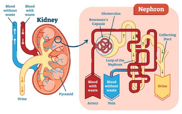
The mechanics and structure of kidneys.
The Project
The goal was to use bioinformatics — making scientific conclusions from large amounts of existing data— to identify which soluble growth factors would be most likely to guide stem cells into becoming kidney cells.
Data was gathered from three databases: the NCBI Gene Database, Pathway Commons, and the R&D Systems. The specific focus was gene expression: how strongly a given gene was being expressed at different stages of development, and if that pattern pointed toward a role in kidney formation.
Nine soluble factors were checked across four criteria — whether they regulate proliferation (division) or apoptosis (cell death), differentiation (specialization), development, and EMT transition where healthy cells become invasive and migratory, as seen in cancer:

Chart of soluble factors’ characteristics.
In addition to the soluble factors themselves, the expression levels of structural proteins were compared across adult kidney tissue and embryonic kidney tissue at 10, 16, and 20 weeks of development. These patterns reveal which proteins the kidney rely on most heavily at each stage, which points towards the growth factors that need to be active during that time.
What the Data Shows
Several patterns emerged from the expression data. Beta-1 integrin (β1) showed the highest expression of all beta subunits in adult kidney tissue at 82.622, declining steadily across embryonic stages. Integrin α6 was notably elevated in both adult and early embryonic kidneys. Laminin subunit LAMB1 peaked in 10-week embryonic tissue at 31.808 before dropping sharply. Fibronectin (FN1) hit 44.16 at 10 weeks of embryonic development — the highest point in its trajectory — and then fell significantly by 20 weeks.
Taken together, these patterns suggest that early kidney development is particularly active in building out the extracellular matrix, the structural scaffolding that surrounds and supports cells. Growth factors that regulate matrix organization are strong candidates for guiding stem cells towards becoming kidney cells. The factors with roles in both differentiation and development — Activin A, Retinoic Acid, and Transforming Growth Factor Alpha — are the most likely contributors based on this analysis.


Data from Bioinformatics Kidney Research
Limitations
The most significant limitation of this project is that the proposed combination of soluble factors was not experimentally tested. The conclusions are based on published data and gene expression databases, not original lab work. The identified factors are candidates, not lab-confirmed drivers of kidney differentiation.
The analysis also focused on general kidney cells. The nephron’s substructures — the glomerulus, Bowman’s capsule, and the loop of the nephron — each contain individual cell types and would their own targeted factor combinations to differentiate properly. That level of specificity is the next step.
Why It Matters
The applications of this research go beyond solving the organ donor shortage. A bioengineered kidney built from a patient’s own iPSCs would carry no foreign genetic material, greatly reducing the risk of immune rejection.
Beyond transplantation, a lab-grown kidney could be used for research. Kidney diseases could be studied in a controlled environment, drug responses tested, and disease mechanisms traced in ways that are not currently possible with donor tissue.
The path from a bioinformatics analysis to a functioning transplantable organ is a long journey, and many technical problems remain unsolved. But identifying the right chemical signals to guide stem cell differentiation is where that path begins.
Conclusion
This project did not produce a kidney. What it produced is a map of the growth factors most likely to be involved in making one. The next step — testing those factors in a lab setting and observing whether the stem cells respond as predicted — is where the bioinformatics becomes bioengineering. If the factors preform as expected, the work of engineering patient-specific kidneys moves one step closer to reality.
References
National Center for Biotechnology Information. (2025). National Library of Medicine. National Institutes of Health. https://www.ncbi.nlm.nih.gov/gene
R&D Systems. (2025). Embryonic and induced pluripotent stem cell differentiation pathways & lineage-specific markers. Bio-Techne. https://www.rndsystems.com/pathways/embryonic-induced-pluripotent-stem-cell-differentiation-pathways-lineage-specific-markers
Kramer, J., Steinhoff, J., Klinger, M., Fricke, L., & Rohwedel, J. (2006). Cells differentiated from mouse embryonic stem cells via embryoid bodies express renal marker molecules. Differentiation, 74(2–3), 91–104. https://doi.org/10.1111/j.1432-0436.2006.00062.x
Xiaohui Ren, Jingya Zhang, Xiaowen Gong, Xin Niu, Xuejin Zhang, Peng Chen, Xuejun Zhang, Differentiation of murine embryonic stem cells toward renal lineages by conditioned medium from ureteric bud cells in vitro, Acta Biochimica et Biophysica Sinica, Volume 42, Issue 7, July 2010, Pages 464–471, https://doi.org/10.1093/abbs/gmq046
Kobayashi, T., Tanaka, H., Kuwana, H., Inoshita, S., Teraoka, H., Sasaki, S., & Terada, Y. (2005). Wnt4-transformed mouse embryonic stem cells differentiate into renal tubular cells. Biochemical and Biophysical Research Communications, 336(2), 585–595. https://doi.org/10.1016/j.bbrc.2005.08.136
Morizane, R., Monkawa, T., & Itoh, H. (2009). Differentiation of murine embryonic stem and induced pluripotent stem cells to renal lineage in vitro. Biochemical and Biophysical Research Communications, 390(4), 1334–1339. https://doi.org/10.1016/j.bbrc.2009.10.148
Dankers, P. Y. W., Boomker, J. M., Meijer, E. W., Popa, E. R., & van Luyn, M. J. A. (2011). From kidney development to drug delivery and tissue engineering strategies in renal regenerative medicine. Journal of Controlled Release, 152(1), 177–185. https://doi.org/10.1016/j.jconrel.2011.01.034
메타데이터
- post_id
- de65a535f4ab
- slug
- bioengineering-stem-cells-for-creating-vital-organs-using-growth-factors-de65a535f4ab
- url
- https://medium.com/@priscilla.suetyau.wong/bioengineering-stem-cells-for-creating-vital-organs-using-growth-factors-de65a535f4ab
- canonical_url
- https://medium.com/@priscilla.suetyau.wong/bioengineering-stem-cells-for-creating-vital-organs-using-growth-factors-de65a535f4ab
- author_url
- https://medium.com/@priscilla.suetyau.wong
- status
- ok
- fetched_at
- 2026-06-09 15:37:30